Urologic Emergencies

The urinary tract is a complex group of organs that includes the kidneys, ureters, bladder, and urethra, plus nerves and muscles. Working together, these components clear waste from the body in the form of urine. For most people, urinating several times a day is part of their daily routine.

There are times, however, when the urinary tract doesn’t work as well as it should. Whether it’s from injury or infection, a person can have a range of symptoms, including pain, trouble with the urine flow, and blood in the urine.

When a person has symptoms like these, it can be hard to know what to do. Should a person wait and see what happens? Or could there be an emergency that needs immediate care?

Woman holding sticky note above her lower abdomen with letters SOS

It’s important to know what constitutes a urologic emergency and how to handle it. This article covers the following urologic emergencies and discusses the definition, symptoms, causes, and treatments for each one. It also explains how one might reduce the risk of such an emergency:

Blood in the urine: A common symptom

As you read about the conditions below, you’ll see that blood in the urine (hematuria) is a common symptom of urologic emergencies. Indeed, it can be upsetting to discover blood in your urine. This situation does not always signify an emergency, but it’s best to have it checked out by a doctor, along with any other symptoms you’re having.

Illustration of urine colors containing increasing amounts of blood ranging from pale yellow to red

Paraphimosis

What is paraphimosis?

Men who are not circumcised still have the foreskin of their penis. Typically, they pull back the foreskin when they clean their penis, urinate, or have sex. The foreskin may also be retracted if a man has a catheter during a surgical procedure.

Paraphimosis happens when the foreskin gets stuck in this retracted position. The man is unable to pull the foreskin forward over the head of the penis.

What are the symptoms of paraphimosis?

In addition to being unable to pull the foreskin forward, men with paraphimosis may have pain, swelling, trouble urinating, and discoloration of the penis (blue or black).

What causes paraphimosis?

Paraphimosis may develop if the penis has been injured. Infection is another cause, especially if a man has poor hygiene. It can also happen if a man does not pull the foreskin forward again after bathing or urinating.

Why is paraphimosis an emergency?

Without treatment, paraphimosis can lead to poor blood flow to the tip of the penis, cutting off the blood and oxygen supply. If this happens, there can be tissue damage or tissue death (gangrene).

How is paraphimosis treated?

Men with paraphimosis should seek treatment immediately.

To start, the doctor will try to reduce swelling at the tip of the penis. This can be done by gently squeezing the area for up to 30 minutes. Sometimes, ice is applied. Once the swelling is reduced, it may be possible to move the foreskin forward.

If this approach isn’t successful, there are other options:

  • Using medications to reduce swelling.
  • Cutting a small slit into the foreskin to loosen it so that it can be pulled forward.
  • Doing a circumcision (surgically removing the foreskin).

If there is an infection, the doctor may prescribe antibiotics.

How might paraphimosis be prevented?

Getting circumcised is the only way to completely prevent paraphimosis. But men can reduce their risk by making sure the foreskin is pulled forward again to cover the tip of the penis after urinating, sex, washing, and medical procedures.


Priapism

What is priapism?

Priapism is an erection that lasts for 4 hours or longer.

Typically, when a man has an erection, his penis fills with blood, which makes it firm. Veins constrict to keep the blood in. After he ejaculates (or when sexual stimulation stops), the veins open again and the blood flows back into the body.

Priapism usually isn’t related to sexual activity.

There are two types of priapism:

  • Ischemic priapism (also called low-flow priapism) occurs when blood cannot flow out of the penis. This is the most common type.
  • Nonischemic priapism (also called high-flow priapism) occurs when more blood than usual flows into the penis.

What are the symptoms of priapism?

The main symptom of priapism is an erection lasting several hours. However, other symptoms can vary depending on the type of priapism.

  • Ischemic priapism is usually painful. The shaft of the penis may be hard, but the tip may be soft.
  • Nonischemic priapism tends to be less painful, but the shaft and tip may remain firm.

What causes priapism?

Priapism can have several causes. Some of the more common ones include:

  • Blood diseases, such as sickle cell anemia or leukemia
  • Side effects to medications, like some erectile dysfunction drugs and antidepressants
  • Trauma to the genital area or spinal cord, such as from an accident
  • Use of alcohol or recreational drugs, such as marijuana or cocaine

Why is priapism an emergency?

When blood is trapped in the penis, it does not receive enough oxygen. If this situation lasts a long time, tissues in the penis can be severely damaged. Erectile dysfunction can also result.

The longer the delay of treatment, the greater the risk of damage.

How is priapism treated?

Doctors make treatment decisions based on the type of priapism a person has.

If a man has ischemic priapism, blood will need to be drained from the penis as soon as possible. Doctors may use one of the following techniques:

  • Aspiration. Blood is drained with a surgical needle and syringe.
  • Injection of medication or saline. This helps blood flow out of the penis.
  • Surgery. Surgical procedures might be done to drain the blood. A shunt may be placed to reroute the blood flow.

Men with nonischemic priapism may not need treatment, as the erection may go away on its own. Ice packs and pressure on the perineum (the area between the anus and the scrotum) might help. If blood vessels have been damaged, they may be repaired with surgery.

Only a doctor can determine the type of priapism, so it’s critical to seek emergency care.

How might priapism be prevented?

Men with blood disorders may reduce their risk for priapism by following the treatment plan prescribed by their doctor. Men should also be careful about the prescription medications they take and limit their use of alcohol and recreational drugs. If priapism is a side effect of medication, the drug may be changed, but this should be done under a doctor’s care.

Decreasing risk of injury, such as wearing seatbelts in the car, may also reduce risk of priapism.


Penile Fracture

What is penile fracture?

Penile fracture refers to a tear in the tunica albuginea, the layer of tissue that coats and protects the corpora cavernosa—two cylinders in the penis that fill with blood during an erection. (Note: There are no actual bones in the penis.)

A fracture happens when there is trauma to the erect penis. For example, if the penis is hit or severely bent, it may fracture.

What are the symptoms of penile fracture?

  • A popping or cracking sounds when the fracture occurs
  • Sudden, intense pain
  • Swelling
  • Bruising
  • Blood at the tip of the penis or in the urine
  • Loss of erection

What causes penile fracture?

Penile fracture can happen:

  • During intercourse.
  • If a man masturbates vigorously.
  • If a man falls on or rolls over on his erect penis.
  • During taqaandan, a cultural practice in which men bend the tip of their penis to stop an erection.

Why is penile fracture an emergency?

Without treatment, a penile fracture can lead to a curved penis or erectile dysfunction (ED). Men may also start having trouble urinating, and scar tissue could form.

How is penile fracture treated?

Men with penile fracture usually have surgery to repair the tunica albuginea.

How might penile fracture be prevented?

Men can reduce their risk for penile fracture by being especially cautious during sex and by not practicing taqaandan.


Fournier’s Gangrene

What is Fournier’s gangrene?

Fournier’s gangrene is a rare, but life-threatening, bacterial infection that can affect the genitals and perineum. (In men, the perineum is the area between the anus and the scrotum. In women, it’s the area between the anus and the vulva.) In more severe cases, it may affect the thighs, stomach, and chest.

If not caught early, Fournier’s gangrene can cause serious tissue damage, organ failure, and death. Patients with this illness may need to spend several weeks in the hospital.

Fournier’s gangrene is 10 times more common in men than in women.

What are the symptoms of Fournier’s gangrene?

A person with Fournier’s gangrene may have the following symptoms:

  • Pain
  • Tenderness and swelling in the genitals or perineum
  • Discolored skin (such as red, purple, brown, or black)
  • Itching
  • Fever and chills
  • Nausea and vomiting
  • Foul odor
  • Mental changes
  • Dehydration
  • Lethargy

What causes Fournier’s gangrene?

Fournier’s gangrene can be caused by several types of bacteria, including E. coli, staphylococcus (“staph”) and streptococcus (“strep”). Sometimes it’s caused by a combination of bacteria.

There are many ways bacteria can enter the body. Examples include genital piercings, scratches or burns on the body, urinary tract infections, insect bites, and sex.

Why is Fournier’s gangrene an emergency?

Fournier’s gangrene is a type of flesh-eating disease. It can quickly destroy blood vessels, muscles, and nerves.

Without prompt treatment, a person can go into septic shock, a life-threatening condition that causes organs to shut down.

How is Fournier’s gangrene treated?

Surgery to remove damaged tissue is often necessary. (Reconstruction surgery to rebuild these areas might occur at a later date.) Antibiotics and other medicines may also be prescribed.

Some patients undergo hyperbaric oxygen therapy, where they inhale pure oxygen. This type of therapy is often associated with scuba divers who get decompression sickness, but it is also used to fight bacteria and heal wounds.

How might Fournier’s gangrene be prevented?

Good hygiene and proper wound care can reduce a person’s risk for Fournier’s gangrene and infections in general. People should also make sure they seek urgent medical care if they notice any redness, tenderness, or swelling in their genitals. For best outcomes, Fournier’s gangrene must be treated as quickly as possible.


Urethral Injuries

What are urethral injuries? What causes them?

The urethra is the tube that allows urine to exit the body. It can be torn or crushed during trauma.

There are several situations that can cause urethral injuries:

  • Trauma, such as from a car accident or serious fall
  • Straddle injuries, when a person is hit hard between the legs
  • Vaginal childbirth
  • Problems with catheters or other urological devices
  • Sexual assault and violence involving weapons
  • Vigorous sexual intercourse

What are the symptoms of urethral injuries?

Common symptoms of urethral injuries include:

  • Pain in the genitals or abdomen
  • Swelling
  • Trouble with urination
  • Blood in the urine (hematuria)
  • Blood at the tip of the penis
  • Bruising on the perineum

Why are urethral injuries an emergency?

Without prompt treatment, urethral injuries can make a person more prone to infections that can affect the urinary tract and kidneys. Urethral injuries may also make it difficult for urine to leave the body, increasing the risk of infections and kidney damage.

Urethral stricture—a narrowing of the urethra—is another potential complication. When this happens, it becomes difficult to empty the bladder.

How are urethral injuries treated?

Urethral injuries are often treated with surgery to repair the tear and other damage. In some cases, a catheter may be placed in the urethra or bladder so that urine can drain while the urethra heals. Patients may receive antibiotics if they have an infection.

How might urethral injuries be prevented?

A person can reduce their risk for urethral injuries by taking the following precautions:

  • Drive safely and wear seat belts.
  • Operate other motor vehicles and machinery safely.
  • Use protective gear, such as athletic supporters, while playing sports.
  • Make sure the home is safe and use assistive devices to prevent falls.
  • Be gentle during sexual activities. Use lubricant.

Testicular Torsion

What is testicular torsion?

Testicular torsion affects the testicles (also called the testes) — two egg-shaped organs found in the scrotum (a sac located beneath the penis). Testicles produce hormones and sperm cells. Each testicle is attached to a spermatic cord, which provides the blood supply needed to keep the testicle healthy.

Sometimes, a testicle and its spermatic cord become twisted, blocking the blood supply to the attached testicle. This is called testicular torsion.

What are the symptoms of testicular torsion?

Men experiencing testicular torsion usually have sudden, intense pain in the testicle. The pain may spread to the abdomen. One testicle may be higher than the other.

There may also be:

  • Swelling
  • Discoloration of the scrotum
  • A lump on the testicle
  • Nausea and vomiting
  • Fever
  • A need to urinate more often

Testicular torsion is possible in both testicles, but it usually affects just one.

What causes testicular torsion?

Testicular torsion usually affects people with a “bell clapper” deformity in their scrotum. Typically, there is tissue that attaches the testicle to the wall of the scrotum, which keeps it from twisting. But some men are born without this attaching tissue, so the testicle swings as a bell clapper would.

Often, there is no specific event that triggers testicular torsion. It can happen while a man is active or sedentary.

Why is testicular torsion an emergency?

The testicle cannot live long without an adequate blood supply. If testicular torsion is not treated within 6 hours, there can be permanent damage to the testicle, and it may need to be surgically removed.

Some men have lower sperm counts after testicular torsion.

How is testicular torsion treated?

Testicular torsion is treated with surgery. The surgeon untwists the spermatic cord so that blood flow can resume. If the testicle can be saved, the surgeon uses stitches to attach it to the scrotum wall, so that it cannot twist again. The other testicle may also be stitched to the scrotum wall to prevent twisting on that side.

If the testicle cannot be saved, it is removed.

How might testicular torsion be prevented?

Men with bell clapper deformity may prevent future episodes of testicular torsion by having their testicle surgically attached to the scrotum wall.


Testicular Rupture

What is testicular rupture?

A man’s two testicles are important for his reproductive health. They produce the hormone testosterone as well as sperm cells.

The testicles are found in the scrotum, a sac near the penis. Each testicle is protected by a membrane called the tunica albuginea. However, the testicles are not protected by muscles or bones, so they can be especially vulnerable to injury.

Testicular rupture occurs when there is forceful trauma to the testicle. Without prompt treatment the testicle can be severely damaged or lost.

What are the symptoms of testicular rupture?

Men feel intense pain when their testicle is ruptured. They may also have:

  • Bruising and swelling in the area
  • Blood in the urine
  • Problems with urination
  • A fever

What causes testicular rupture?

Testicular rupture is caused by blunt force or piercing injuries, such as the following:

  • Vehicular accidents
  • Falls
  • Injuries while playing sports
  • Violence involving knives or guns

Why is testicular rupture an emergency?

If a testicular rupture is not treated quickly, there is a greater risk of infection. Blood supply to the testicle can also be compromised. Without good blood supply, the testicle cannot survive.

How is testicular rupture treated?

Men with testicular rupture usually have surgery to repair it. If the rupture is severe, the testicle may need to be removed.

How might testicular rupture be prevented?

Men can reduce their risk for testicular rupture by taking these precautions:

  • Drive safely and wear seat belts.
  • Operate other motor vehicles and machinery safely.
  • Use protective gear, such as athletic supporters, while playing sports.
  • Being careful to avoid injury to the genital area.

Acute Kidney Injury

What is acute kidney injury?

Acute kidney injury (AKI) is a sudden instance of kidney failure. When AKI happens, the kidneys can no longer filter the blood effectively. As a result, waste products like creatinine and urea accumulate in the blood.

AKI usually begins over the course of a few hours or days. It often affects older people and patients who are already in the hospital for another illness. Chronic kidney disease, diabetes, and dehydration can increase a person’s risk for AKI.

When it is severe, it can be life-threatening.

An older term for AKI is acute renal failure.

What are the symptoms of acute kidney injury?

Many people don’t have symptoms until the later stages of AKI. However, symptoms typically include:

  • Passing less urine than usual
  • Swelling
  • Fatigue
  • Nausea
  • Shortness of breath
  • Chest pain
  • Confusion
  • Seizures or coma

What causes acute kidney injury?

There are many conditions and situations that can lead to AKI. Here are some examples:

  • Conditions that decrease blood flow to the kidneys, such as low blood pressure, severe diarrhea, heart failure, and organ failure
  • Conditions that cause kidney damage, such as sepsis, multiple myeloma, vasculitis, and interstitial nephritis
  • Overuse of nonsteroidal anti-inflammatory drugs (NSAIDs), such as ibuprofen and naproxen

Why is acute kidney injury an emergency?

Without treatment, AKI can lead to permanent kidney damage and, in rare cases, death. It can also upset the balance of fluids and electrolytes in the body as waste products accumulate in the blood.

How is acute kidney injury treated?

Doctors treat AKI by treating the condition that causes it. For example, if medication is the cause, the doctor may prescribe alternatives. If there is an infection, then that will be treated.

In severe cases, patients with AKI are put on dialysis. Dialysis is a procedure that filters the blood when the kidneys are unable to do so.

How might acute kidney injury be prevented?

People can lower their risk for AKI by paying attention to their kidney health and managing conditions that are risk factors, like dehydration, diabetes, high blood pressure, and heart disease.


Obstructed Pyelonephritis (Pyonephrosis)

What is obstructed pyelonephritis?

The word pyelonephritis refers to a kidney infection. A person with obstructed pyelonephritis has a kidney infection along with a blockage in the urinary tract.

What are the symptoms of obstructed pyelonephritis?

Symptoms of obstructed pyelonephritis tend to come on suddenly, over the course of a few hours or within a day. They may include:

  • Pain in the lower back or flank
  • Pain or burning sensation during urination
  • Fever or chills
  • Nausea or vomiting
  • Bloody urine
  • Needing to urinate more frequently or urgently
  • Releasing less urine than usual
  • Confusion or changes in mental state
  • Shortness of breath

What causes obstructed pyelonephritis?

Most of the time, obstructed pyelonephritis is caused by a bacteria, although sometimes it’s caused by a virus. The bacteria typically enter the body through the urethra, then travel up the urinary tract.

In addition to the bacteria, there is a blockage in the urinary tract. This obstruction might be caused by kidney stones, an enlarged prostate, uterine prolapse, or another condition. Because the blockage makes it harder for urine to leave the body, bacteria can grow in the backed-up urine.

Why is obstructed pyelonephritis an emergency?

Without treatment, obstructed pyelonephritis can lead to kidney failure, kidney damage, abscesses, sepsis, or blood clots in the veins of the kidney.

How is obstructed pyelonephritis treated?

Obstructed pyelonephritis is treated with antibiotics and other medicines. Some patients need to be hospitalized. In some cases, surgical drainage may be needed.

How might obstructed pyelonephritis be prevented?

People might lower their risk for obstructed pyelonephritis by:

  • Staying properly hydrated.
  • Emptying the bladder whenever they urinate.
  • Wiping from front to back after using the bathroom.
  • Urinating before and after sexual activity.
  • Keeping the genital area clean.

Bladder Injury

What is a bladder injury? What causes it?

The bladder is the organ that stores urine after it is produced by the kidneys. It can be damaged through blunt force (such as in a motor vehicle accident or while playing sports). It may also be torn by penetrating injuries (such as knife or gunshot wounds).

What are the symptoms of a bladder injury?

A person with a bladder injury may have symptoms like these:

  • Pain in the abdomen or back
  • Blood in the urine
  • Blood or urine coming from the vagina.
  • Difficulty with urination or inability to urinate at all
  • Painful or frequent urination
  • Fever
  • Bruising

Why is a bladder injury an emergency?

Without prompt treatment, a bladder injury can have complications such as:

  • Sepsis
  • Infections
  • Kidney problems
  • Permanent bladder damage
  • Urinary incontinence
  • Fistulas (connections between two organs that don’t typically connect, such as the bladder and the intestines)

How is a bladder injury treated?

Treatments for bladder injuries depend on the severity of the injury.

In mild cases, patients may have a temporary catheter to drain urine out of the body. This gives the bladder a chance to heal.

More serious cases may require surgery to repair the tear or other damage.

Medications to manage pain and treat any infections may also be prescribed.

How might a bladder injury be prevented?

People may lower their risk of bladder injuries by following safety precautions when operating motor vehicles and playing sports.


Acute Urinary Retention

What is acute urinary retention? What are the symptoms?

Acute urinary retention occurs when a person is unable to urinate, even if they have a full bladder. (In some cases, people may urinate only a very small amount.) It comes on suddenly and can be quite painful. People may have abdominal swelling and still feel the need to empty their bladder.

This condition is different from chronic urinary retention, in which a person can still urinate, but their bladder doesn’t empty. Chronic urinary retention occurs gradually and is not an emergency.

Acute urinary retention is more frequent in older men, and the risk increases with age. Women and children are less likely to develop acute urinary retention.

What causes acute urinary retention?

In men, acute urinary retention is often caused by an enlarged prostate gland. The prostate is a walnut-sized gland in a man’s reproductive system. The urethra (the tube that allows urine to exit the body) runs right through the middle of the prostate.

As men get older, their prostate tends to get larger. However, the direction of this growth is inward rather than outward. As a result, excess prostate tissue can squeeze the urethra, making urination more difficult. And in some cases, the growth can block urine flow completely.

Other causes of acute urinary retention may include:

  • Urethral stricture (narrowing of the urethra)
  • Urinary stones
  • Side effects of medications, such as some antihistamines and antidepressants
  • Nerve issues
  • Swelling from a prostate infection or urinary tract infection

Why is acute urinary retention an emergency?

Without treatment, acute urinary retention can lead to bladder and kidney damage. Bacteria that remain in the bladder can cause a urinary tract infection.

How is acute urinary retention treated?

In the emergency department, the doctor will insert a urinary catheter into the bladder to drain it.

After the initial emergency is addressed, the doctor may suggest treatments for the underlying cause of the retention. For example, an enlarged prostate may be treated with drugs or medical procedures. Urethral stricture may be treated with surgery. If medications are the cause, the doctor may adjust the dose or change the prescribed drug.

How might acute urinary retention be prevented?

People might lower their risk for acute urinary retention by:

  • Not holding their urine
  • Staying hydrated
  • Getting enough fiber in their diet
  • Exercising regularly
  • Doing pelvic floor exercises
  • Taking medicine as prescribed

Kidney Stones

What are kidney stones? What causes them?

Kidney stones are small masses that form in your kidney. They form from a buildup of substances, like calcium and uric acid, that crystallize and form stones. Some stones pass on their own, but others get stuck in the urinary tract.

What are the symptoms of kidney stones?

Some of the common symptoms of kidney stones are:

  • Pain
  • Blood or “sand” in the urine (Sand refers to tiny stones that pass in the urine, which resemble sand or gravel.)
  • Changes in urination
  • Nausea
  • Fever or chills

Why might kidney stones be an emergency?

Kidney stones are not always an emergency. In fact, some people with kidney stones have no symptoms at all. Others have mild symptoms that can be managed with medication at home until the stones pass.

However, for some people, the pain of kidney stones is intense. Also, the size of the stone and the potential for hydronephrosis (swelling of the kidney) can be concerns. In those cases, going to an urgent care clinic or emergency room is recommended.

How are kidney stones treated?

Severe kidney stones can be treated in several ways:

  • Ureteroscopy. A thin, long tube is threaded through the urinary tract and into the kidney. This tool can remove stones or break them down to be passed through urine.
  • Shock wave lithotripsy (SWL). Shock waves, administered through the skin, break down the stones so they can pass.
  • Percutaneous nephrolithotomy (PNL). Stones are surgically removed through a small incision in your back or side.

Kidney stones are not always an emergency. However, for some people, the pain of kidney stones is intense.

How might kidney stones be prevented?

You can lower your risk for kidney stones by staying hydrated (drinking plenty of water). If you have had stones before, your doctor may recommend a special diet or taking prescribed medications.

Learn more about kidney stones.


Resources

Paraphimosis

American Academy of Family Physicians

“Paraphimosis”
(Last Updated: January 2021)
https://familydoctor.org/condition/paraphimosis/

Cleveland Clinic

“Paraphimosis”
(Last review: March 19, 2025)
https://my.clevelandclinic.org/health/diseases/22244-paraphimosis

Medical News Today

Ingleson, Kanna
“What to know about paraphimosis”
(August 8, 2017)
https://www.medicalnewstoday.com/articles/318833

MedlinePlus

“Paraphimosis”
(Reviewed: January 1, 2023)
https://medlineplus.gov/ency/article/001281.htm

Up to Date

Diamond, David A, MD and Tews, Matthew, DO
“Paraphimosis: Clinical manifestations, diagnosis, and treatment”
(Topic last updated: August 28, 2024)
https://www.uptodate.com/contents/paraphimosis-clinical-manifestations-diagnosis-and-treatment

Priapism

American Urological Association

Bivalacqua T.J., Allen B.K., Brock G.B., et al.
“Diagnosis and Management of Priapism: AUA/SMSNA Guideline (2022)”
(2022)
https://www.auanet.org/guidelines-and-quality/guidelines/diagnosis-and-management-of-priapism-aua/smsna-guideline-(2022)

Mayo Clinic

“Priapism – Diagnosis and Treatment”
(August 31, 2021)
https://www.mayoclinic.org/diseases-conditions/priapism/diagnosis-treatment/drc-20352010

“Priapism – Symptoms & Causes”
(August 31, 2021)
https://www.mayoclinic.org/diseases-conditions/priapism/symptoms-causes/syc-20352005

Up to Date

Deveci, Serkan, MD
“Priapism”
(Topic last updated: February 10, 2025)
https://www.uptodate.com/contents/priapism

Urology Care Foundation

“What is Priapism?”
(Updated: August 2022)
https://www.urologyhealth.org/urology-a-z/p/priapism

Penile Fracture

American Urological Association

Morey A.F., Brandes S,. Dugi D.D. 3rd, et al.
“Urotrauma (2020)”
(Published: 2014. Amended: 2017 and 2020)
https://www.auanet.org/guidelines-and-quality/guidelines/urotrauma-guideline

Cleveland Clinic

“Penile Fracture”
(Last reviewed: June 7, 2024)
https://my.clevelandclinic.org/health/diseases/21707-penile-fracture

Mayo Clinic

Ziegelmann, Matthew (Matt) J., M.D.
“Is it possible to ”break” your penis?”
(July 6, 2024)
https://www.mayoclinic.org/healthy-lifestyle/sexual-health/expert-answers/penis-fracture/faq-20058154

Up to Date

Voelzke, Bryan, MD, MS, FACS
“Traumatic injury to the male anterior urethra, scrotum, and penis”
(Topic last updated: November 15, 2024)
https://www.uptodate.com/contents/traumatic-injury-to-the-male-anterior-urethra-scrotum-and-penis

Fournier’s Gangrene

Cleveland Clinic

“Fournier’s Gangrene”
(Last reviewed: February 17, 2025)
https://my.clevelandclinic.org/health/diseases/22025-fourniers-gangrene

“Septic Shock”
(Last reviewed: June 14, 2022)
Septic Shock: Causes, Symptoms & Treatment

DermNetNZ.com

Singh, Amritpreet and Amanda Oakley
“Fournier gangrene”
(Copy edited: March 2022)
https://dermnetnz.org/topics/fournier-gangrene

Mayo Clinic

“Hyperbaric oxygen therapy”
(December 6, 2024)
https://www.mayoclinic.org/tests-procedures/hyperbaric-oxygen-therapy/about/pac-20394380

Urethral Injuries

American Urological Association

Morey A.F., Brandes S,. Dugi D.D. 3rd, et al.
“Urotrauma (2020)”
(Published: 2014. Amended: 2017 and 2020)
https://www.auanet.org/guidelines-and-quality/guidelines/urotrauma-guideline

StatPearls via NIH National Library of Medicine

Leslie, Stephen W., et al.
“Urethral Injury”
(Last update: July 24, 2023)
https://www.ncbi.nlm.nih.gov/books/NBK554575/

Urology Care Foundation

“Urethral Trauma”
(no date)
https://www.urologyhealth.org/urology-a-z/u/urethral-trauma

Testicular Torsion

Cleveland Clinic

“Testicular Torsion”
(Last reviewed: February 27, 2023)
https://my.clevelandclinic.org/health/diseases/15382-testicular-torsion

MedlinePlus

“Testicular torsion”
(Review date: July 1, 2023)
https://medlineplus.gov/ency/article/000517.htm

Up to Date

Eyre, Robert C., MD and Farooq, Ahmer, DO
“Acute scrotal pain in adults: Evaluation and management of major causes”
(Topic last updated: April 22, 2025)
https://www.uptodate.com/contents/acute-scrotal-pain-in-adults-evaluation-and-management-of-major-causes

Urology Care Foundation

“Testicular Torsion”
(Updated March 2024)
https://www.urologyhealth.org/urologic-conditions/testicular-torsion

Testicular Rupture

Cleveland Clinic

“Ruptured Testicle”
(Last reviewed: June 6, 2022)
https://my.clevelandclinic.org/health/diseases/23245-ruptured-testicle

Up to Date

Runyon, Michael S., MD, MPH
“Blunt genitourinary trauma: Initial evaluation and management”
(Topic last updated: March 8, 2023)
https://www.uptodate.com/contents/blunt-genitourinary-trauma-initial-evaluation-and-management

Urology Care Foundation

“Testicular Trauma”
(No date)
https://www.urologyhealth.org/urology-a-z/t/testicular-trauma

Acute Kidney Injury

National Kidney Foundation

“Acute Kidney Injury (AKI)”
(Last updated: February 26, 2024)
https://www.kidney.org/kidney-topics/acute-kidney-injury-aki

Yale Medicine

“Acute Kidney Injury (AKI)”
(No date)
https://www.yalemedicine.org/conditions/acute-kidney-injury

Obstructed Pyelonephritis

Cleveland Clinic

“Kidney Infection (Pyelonephritis)”
(Last reviewed: January 31, 2023)
https://my.clevelandclinic.org/health/diseases/15456-kidney-infection-pyelonephritis

StatPearls via National Library of Medicine

Belyayeva, Mariya; Leslie, Stephen W.; and Jeong, Jordan M.
“Acute Pyelonephritis”
(Last update: February 28, 2024)
https://pubmed.ncbi.nlm.nih.gov/30137822/

Bladder Injury

American Urological Association

Morey A.F., Brandes S,. Dugi D.D. 3rd, et al.
“Urotrauma (2020)”
(Published: 2014. Amended: 2017 and 2020)
https://www.auanet.org/guidelines-and-quality/guidelines/urotrauma-guideline

StatPearls via National Library of Medicine

Kang, Lorna and Geube, Aleksandar
“Bladder Trauma”
(Last update: May 22, 2023)
https://www.ncbi.nlm.nih.gov/books/NBK557875/

Urology Care Foundation

“Bladder Trauma”
(No date)
https://www.urologyhealth.org/urology-a-z/b/bladder-trauma

Acute Urinary Retention

Cleveland Clinic

“Urinary Retention”
(Last reviewed: January 23, 2024)
https://my.clevelandclinic.org/health/diseases/15427-urinary-retention

National Institute of Diabetes and Digestive and Kidney Diseases

“Definition & Facts of Urinary Retention”
(Last reviewed: December 2019)
https://www.niddk.nih.gov/health-information/urologic-diseases/urinary-retention/definition-facts

“Preventing Urinary Retention”
(Last reviewed: December 2019)
https://www.niddk.nih.gov/health-information/urologic-diseases/urinary-retention/prevention

“Symptoms & Causes of Urinary Retention”
(Last reviewed: December 2019)
https://www.niddk.nih.gov/health-information/urologic-diseases/urinary-retention/symptoms-causes

“Treatment of Urinary Retention”
(Last reviewed: December 2019)
https://www.niddk.nih.gov/health-information/urologic-diseases/urinary-retention/treatment

Up to Date

Barrisford, Glen W., MD, MS, MPH, FACS and Steele, Graeme S. MBBCh, FCS, FACS
“Acute urinary retention”
(Topic last updated: September 9, 2024)
https://www.uptodate.com/contents/acute-urinary-retention

Kidney Stones

American Urological Association

“Medical Management of Kidney Stones (2019)”

(Published 2014. Reviewed and validity confirmed 2019)

https://www.auanet.org/guidelines/kidney-stones-medical-mangement-guideline

“Surgical Management of Stones: AUA/Endourology Society Guideline (2016)”

(Published 2016)

https://www.auanet.org/guidelines/kidney-stones-surgical-management-guideline

Up to Date

“Patient education: Hydronephrosis in adults (The Basics)”
(no date)
https://www.uptodate.com/contents/hydronephrosis-in-adults-the-basics

Preminger, Glenn M., MD and Gary C. Curhan, MD, ScD
“Patient education: Kidney stones in adults (Beyond the Basics)”
(Last updated: May 16, 2023)
https://www.uptodate.com/contents/kidney-stones-in-adults-beyond-the-basics

Urology Care Foundation

“What are Kidney Stones?”
(no date)
https://www.urologyhealth.org/urology-a-z/k/kidney-stones


Pregnancy and Kidney Stones

What You Should Know About Kidney Stones During Pregnancy

Kidney stones are a fairly common urological problem, affecting about 1 in 10 people at some point in their life. But kidney stones can be a particular challenge for pregnant women. The way stones are diagnosed and treated may need to be adjusted during pregnancy for the safety of the mother and her baby.

Still, pregnant women with kidney stones have several treatment options. And it’s important to know that without treatment, kidney stones can lead to premature labor, so it’s necessary to address them.

Backache at the end of pregnancy

What are kidney stones?

Kidney stones are small, crystallized masses that form in the kidneys or ureters (the tubes connecting the kidneys to the bladder). Most stones are made from calcium, but they can also be formed from uric acid, struvite, and cystine, which are naturally occurring compounds in the human body. Kidney stones come in various shapes and sizes and can be smooth or jagged, small as a pea, or as large as a golf ball.

Most kidney stones are small enough to pass through the urinary tract on their own. A person with a small kidney stone might not have any symptoms.

But larger stones can block urine flow and cause a significant amount of pain. In fact, abdominal pain is one of the most common symptoms of kidney stones. Some people have nausea and vomiting, along with a need to urinate more urgently and frequently. Blood in the urine is another symptom.

How common are kidney stones in pregnant women?

It’s estimated that kidney stones occur in about 1 of every 500 to 3,000 pregnancies, so having kidney stones during pregnancy is fairly rare.

However, research suggests that risk for first-time kidney stone formation is higher at certain points in a pregnancy.

In 2021, the American Journal of Kidney Diseases published a study on the topic. Researchers found that in pregnant women, risk for developing first-time symptomatic kidney stones increased during the second and third trimesters of pregnancy.

Interestingly, the peak time to develop kidney stones was during the first 3 months after delivery. About a year after the delivery date, risk returned to pre-pregnancy levels.

Risk was higher in pregnant women who were obese.

Why are pregnant women more at risk for kidney stones?

Pregnant women are more prone to kidney stones for the following reasons:

  • Pregnant women are more susceptible to dehydration. A growing baby puts pressure on a woman’s bladder, increasing the need to urinate. As a result, pregnant women might not drink as much fluid as they should, and dehydration can lead to kidney stones.
  • Changes occur in urine composition during pregnancy. Pregnant people tend to have more calcium in their urine, and most kidney stones are calcium-based.
  • Hormonal changes in pregnant women make it more difficult to clear urine from the body. Pregnant women have higher levels of the hormone progesterone, which contributes to urinary stasis (halting of or slowing of urine flow). As a result, stone-forming compounds have more opportunity to crystallize.

How are kidney stones diagnosed in pregnant women?

Diagnosis starts with a medical history. Designing a personalized treatment plan requires knowing more about your symptoms, your pregnancy, and your medical background.

Lab tests are also an important diagnostic tool. These include blood tests and urine tests. Urine tests may reveal if stone-forming substances are present.

Imaging tests may also be ordered. These tests can reveal the location, size, and shape of any kidney stones:

  • Ultrasound. Ultrasound technology uses sound waves to create images of internal organs. (It’s also used to monitor the development of a baby in the uterus.) No radiation is used with ultrasounds, so they are perfectly safe for both the patient and the fetus. For a kidney stones diagnosis, ultrasound technicians focus on the kidney and pelvic area.
  • Magnetic resonance (MR) urography. Urography refers to imaging of the urinary tract: kidneys, ureters, bladder, and urethra. Magnetic resonance (MR) technology creates images using magnet and radio waves. Like ultrasound technology, MR does not use radiation.
  • Low-dose computed tomography (CT scan). A CT scan (sometimes called a “cat scan”) uses x-rays to create images. Because this method does involve radiation, it is recommended for the second and third trimesters only, not the first trimester.

How are kidney stones treated in pregnant women?

Kidney stone treatment in pregnant women requires a team approach. Urologists work with obstetricians and other healthcare specialists to make sure treatment is safe and appropriate.

Initial therapies may include the following:

  • Observation. Generally, treatment starts with observation, a “wait and see” approach, as most stones pass on their own. Some doctors recommend bed rest, increased fluid intake, and a low-salt diet.
  • Pain relief. Patients might be given pain relievers, such as acetaminophen. Other medications, such as non-steroidal anti-inflammatory drugs (NSAIDS) might be prescribed depending on the stage of the pregnancy. Patients should take their medication exactly as prescribed.
  • Hydration. Patients may be advised to drink more fluids. Others may receive hydration via intravenous (IV) fluids at the doctor’s office to help stones pass.

If stones do not pass easily, procedures to extract stones or effect passage of urine around stones are considered. If symptoms are not severe, such procedures might be delayed until after the baby is born. In the case of more serious symptoms, trouble urinating, or a urinary tract infection, the patient may undergo treatment during the pregnancy. (In emergencies, these procedures might take place right away.)

The treatment team will carefully consider choices related to any anesthesia and related drugs given during these procedures.
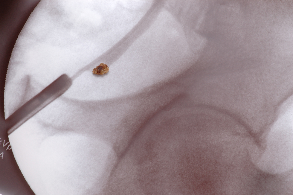
Procedures to extract stones or effect passage of urine around stones include the following:

  • Ureteroscopy. Patients undergoing ureteroscopy receive anesthesia. Once it has taken effect, the urologist places a long, thin instrument called a ureteroscope through the urethra and bladder to the ureter or kidney. The ureteroscope is like a tiny telescope that allows the doctor to see into the affected area. The ureteroscope also has a grasping mechanism that allows it to either remove the stone or break it into smaller stones that can pass through urine.
  • Stent. A stent is a plastic tube that is surgically placed in the ureter to keep it open. With a stent, urine—and the stone—may flow through the ureter more easily. Stents are temporary; in pregnant patients, they are usually replaced every 4 to 6 weeks.
  • Nephrostomy tube. A nephrostomy tube is a type of catheter used to drain urine from the kidney. Typically, urine flows from a kidney to the bladder through a ureter that connects these two organs. With a nephrostomy tube, urine bypasses the ureter and bladder. The surgeon creates a special opening called a stoma on the patient’s side. The nephrostomy tube runs from the kidney and through the stoma, connecting to a urine collection bag outside the body.

    Like stents, nephrostomy tubes are temporary and may need to be changed periodically. Patients receive thorough instructions on the care of their tube and the changing of their urine collection bag.

Resources

American Academy of Family Physicians

“Magnetic Resonance Imaging (MRI)”
(Last updated: June 23, 2020)
https://familydoctor.org/magnetic-resonance-imaging-mri/

American Journal of Kidney Diseases

Thongprayoon, Charat, et al.
“Risk of Symptomatic Kidney Stones During and After Pregnancy”
(Published: April 15, 2021)
https://www.ajkd.org/article/S0272-6386(21)00402-9/fulltext

American Urological Association

Assimos, Dean, et al.
“Surgical Management of Stones: AUA/Endourology Society Guideline (2016)”
(Published: 2016)
https://www.auanet.org/guidelines/guidelines/kidney-stones-surgical-management-guideline

Healthline.com

Seladi-Schulman, Jill, PhD
“Caring for Your Nephrostomy Tube”
(Update: September 18, 2018)
https://www.healthline.com/health/nephrostomy-tube-care

MedlinePlus.gov

“CT Scans”
(Page last updated: April 12, 2021)
https://medlineplus.gov/ctscans.html

“Ultrasound”
(Page last reviewed: December 15, 2020)
https://medlineplus.gov/lab-tests/sonogram/

Medscape

Harrison, Pam
“Pregnancy Increases Risk for Symptomatic Kidney Stone”
(April 28, 2021)
https://www.medscape.com/viewarticle/950073

Nature Reviews | Urology

Semins, Michelle J. and Brian R. Matlaga
“Kidney stones during pregnancy”
(Published online: February 11, 2014)https://www.nature.com/articles/nrurol.2014.17″ https://www.nature.com/articles/nrurol.2014.17

Accessed via: https://svmi.web.ve/wh/intertips/7.UROLITIASIS-Y-EMBARAZO.pdf” https://svmi.web.ve/wh/intertips/7.UROLITIASIS-Y-EMBARAZO.pdf

NEJM Journal Watch

O’Dwyer, Marie Claire, MB BCh BAO, MPH
“Are Pregnant people More Likely to Develop Kidney Stones?”
(September 22, 2021)
https://www.jwatch.org/na54060/2021/09/22/are-pregnant-women-more-likely-develop-kidney-stones

UpToDate

Preminger, Glenn M., MD and Gary C. Curhan, MD, ScD
“Kidney stones in adults: Kidney stones during pregnancy”
(Topic last updated: November 11, 2021)
https://www.uptodate.com/contents/kidney-stones-in-adults-kidney-stones-during-pregnancy

Urology Care Foundation

“Pregnancy and Kidney Stones”
(Summer 2019)
https://www.urologyhealth.org/healthy-living/urologyhealth-extra/magazine-archives/summer-2019/did-you-know-pregnancy-and-kidney-stones

“Preventing and Treating Kidney Stones”
(Summer 2014)
https://www.urologyhealth.org/healthy-living/urologyhealth-extra/magazine-archives/summer-2014/preventing-and-treating-kidney-stones

Urology Marketing Center

“Kidney Stones”
https://www.hsadeghi.com/patient-education/kidney-stones/


Kidney Stones

What are kidney stones? How do they form?

A kidney stone is a small mass that forms from crystallized substances in your kidney or, in some cases, one of your ureters (the tubes that connect the kidneys to the bladder). These chemicals aren’t able to dissolve in urine, so the crystals stick together and get larger, forming the stone. (Note: Stones that form in the ureters are called ureteral stones.)

Abstract image of kidney disease and kidney stones

You can be especially prone to kidney stones if you’re frequently dehydrated and there is less water to dissolve the substances. You can also develop them if your levels of the chemicals that crystallize to form stones are too high.

Kidney stones vary in size, shape, and color.
They are also made from different substances, depending on the type of stone.

Stones vary in texture and size. Some are smooth, while others have more jagged edges. Most stones are less than 5 mm in diameter (about 3/16 of an inch) and they usually pass on their own through urine. But sometimes, stones become larger (even as large as a golf ball) or have trouble passing through the urinary tract. If this happens, you’ll need treatment, which we’ll discuss below.

Other terms for kidney stones are renal stone disease, nephrolithiasis and urolithiasis. Why are there so many names for kidney stones? The other names are scientific names. Both the word “renal” and the prefix “nephro” mean “relating to the kidneys” and scientists may sometimes refer to kidney stones as renal calculi or nephroliths.

Who gets kidney stones?

The American Kidney Fund estimates that about 1 in 5 males and 1 in 14 females will get a kidney stone in their lifetime. Each year, about half a million people go to the emergency room because of kidney stones, the National Kidney Foundation reports.

People of all ages can get kidney stones.

What causes kidney stones?

Kidney stones form from substances found in your urine, like uric acid, calcium, oxalate, and cystine. You can get kidney stones if you have high levels of these substances in your system. But you might also get them if your body doesn’t make enough urine.

In either case, tiny crystals form and gradually get bigger, making a stone. Most of the time, stones just travel through your urinary tract and pass with your urine. But sometimes, they get stuck on the way and block urine flow.

What are the risk factors for kidney stones?

Here are some common risk factors for kidney stones:

Your previous health history. If you have had kidney stones in the past or if you’ve had gastric bypass surgery, you could be at higher risk.

Your family history. If you have an immediate family member – a parent, brother or sister – with a history of kidney stones, you’ll be more likely to develop them yourself.

Dehydration. When you become dehydrated, your body doesn’t produce as much urine and stone-forming substances have a harder time dissolving.

Bowel problems. Frequent diarrhea can make you dehydrated.

Your diet. High levels of calcium, salt, sugar, and animal protein can make you more susceptible to kidney stones. It’s possible that you’re consuming too much of these items in your diet. But it’s also possible that your body isn’t processing them as it should. Once we know more about your kidney stones, we can advise you on foods to choose and foods to avoid.

Health conditions. Obesity, abnormal growth of parathyroid glands (glands that produce a hormone that regulates levels of calcium and phosphorus in the body), and distal renal tubular acidosis (excess amounts of acid in the blood as a result of poor kidney function) are kidney stone risk factors.
Other health issues that can raise your risk for kidney stones are diabetes, polycystic kidney disease, arthritis, and gout.

Medications and supplements. For example, if you take calcium or vitamin C supplements, you might be more prone to kidney stones. If you make a list of the medications and supplements you take, we can review it with you.

How are kidney stones classified?

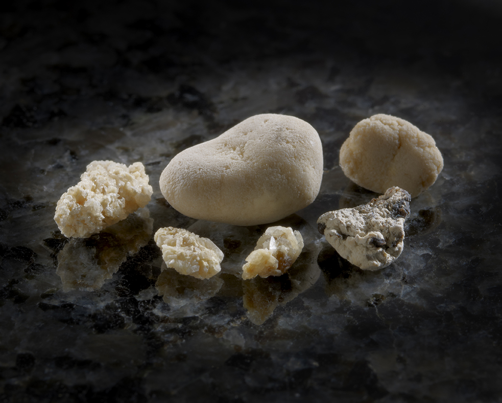
Six kidney stones in various sizes and textures
These kidney stones are all from the same person, but on different occasions. The bigger is 11 mm across. All came out the natural way, without surgery.

Kidney stones vary in size, shape, and color. They are also made from different substances, depending on the type of stone. Knowing of the type of kidney stone you have helps us treat it most effectively.

Calcium stones. Most kidney stones (about 80%) are made from either calcium oxalate or calcium phosphate. These can form if there’s too much calcium in your urine.

Uric acid stones. Uric acid is produced when your body breaks down compounds called purines, which are found in foods like dried beans, peas, and some meats and shellfish. When uric acid crystals accumulate, they can form a uric acid stone.

Struvite stones. Struvite stones (sometimes called “infection stones”) are more common in people who get frequent urinary tract infections (UTIs). Bacteria that cause UTIs disrupt the chemical balance in the urine. Struvite stones are sometimes called staghorns because they have a jagged appearance.

Cystine stones. This type of stone, made from an amino acid called cystine, is caused by a rare inherited condition called cystinuria. People with cystinuria have too much cystine in their urine.

Symptoms

Symptoms may be different depending on the type of stone you have, but some of the more typical symptoms are:

Back pain, kidney inflammation, man suffering from backache at home

Pain

You might have heard stories about pain associated with kidney stones because the pain can be intense, but it’s not always so. Usually pain occurs when urine can’t flow past the stone. You might feel pain in your back, side, abdomen, or groin. Pain can be mild or severe, and it often fluctuates.

Periods of severe pain are called renal colic. These episodes can last up to an hour.

We can help you manage the pain, but if it becomes unbearable and you are unable to reach us, don’t hesitate to head to the ER.

Some men feel pain in their testes or penis as the stone passes. Once the stone is passed, the pain stops.”

Blood in your urine (hematuria) or “sand.”

If you have blood in your urine, you might see a pink or reddish tint in the toilet. But it’s also possible that you won’t see anything different at all. Microhematuria (microscopic hematuria) occurs when the blood particles are too small to see with the naked eye.

You might also notice tiny stones in your urine. Urologists call the particles “gravel” or “sand.”

Urination changes

You might urinate more frequently or feel like you need to go to the bathroom more often, even if you don’t produce much or any urine. You could also have pain or a burning sensation when you urinate. Urine may be cloudy or have an odor.

Nausea. You might start vomiting.

Fever and chills. You may have these symptoms if your stone is related to a UTI.

It’s also possible for people with kidney stones to have no symptoms at all. In fact, some people have kidney stones for years without experiencing any problems.

Diagnosing kidney stones

When you come see us, we’ll start by taking a complete medical history. Be sure to tell us about your diet. That can give us some important clues. We’ll also ask about the medications you take and whether anyone in your family has had stones before.

Next, we’ll do some tests, which may include the following:

  • Urine tests and blood tests. Substances that form stones, like calcium and uric acid, may be present in your urine or blood.
  • Imaging tests. A CT scan (a “cat” scan – computed tomography) or an ultrasound can indicate the size, shape, and quantity of kidney stones.
  • Stone analysis. If you’ve had kidney stones before (and one has been saved), analyzing a previous stone might provide clues for your current situation.
  • Genetic tests. Some kidney stones are caused by inherited conditions, like cystinuria. Genetic tests might tell us if you are likely to develop kidney stones.

Kidney stone treatment

If your stone is small and your pain is mild, you might be able to manage your kidney stone at home. If it’s safe for you, we’ll recommend nonsteroidal anti-inflammatory drugs (NSAIDS) like ibuprofen or naproxen. We might also prescribe medication like tamsulosin (Flomax), which can help your stone pass by relaxing the ureter. This process can take a few weeks.

We might ask you to strain your urine when you’re home. If you catch the stone as it passes, save it and bring it to us for analysis. This will help us determine what type of stone it is.

If your stone is large or doesn’t pass on its own after 4 to 6 weeks, you’ll need more advanced treatment. You will likely have imaging tests and a urinalysis beforehand. These tests results help us choose the best treatment for you.

Ureteroscopy

This procedure is generally used to treat stones in your kidney or ureter. After you have received anesthesia, a thin tool called a ureteroscope is threaded through your urethra and bladder until it reaches the ureter or kidney. The ureteroscope offers doctors a better view of the stone. It can also remove the stone or break it into smaller pieces that can pass on their own.

Chances are, you’ll be able to go home the same day as your ureteroscopy and go back to your daily routine in two to three days.

After a ureteroscopy, you might have a temporary stent — a small tubelike structure — placed in your ureter to keep it open. The stent is usually removed about four to ten days after your procedure.

Shock wave lithotripsy (SWL)

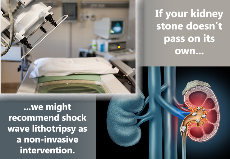
SWL uses shock waves that go through your skin, hit the stone’s surface, and break it into smaller pieces that can pass more easily. You will likely receive anesthesia. Many patients go home the same day as their procedure and can resume their normal activities in two or three days. Afterward, you might see blood in your urine, and you could still have some discomfort while the remaining kidney stone fragments pass. We might prescribe medication to help them pass.

SWL isn’t recommended for people with large or hard stones.

If your kidney stone doesn't pass on its own, we might recommend shock wave lithotripsy as a non-invasive intervention. Accompanying issustration of kidney with stones and photo of shock wave lithotripsy machine.

Percutaneous nephrolithotomy (PNL)

Illustration of remove kidney stones

PNL can be helpful for people with larger kidney stones. You will be anesthetized for the procedure. A small incision will be made in your back or side. This incision allows doctors to examine your kidney with a tool called a nephroscope. Another instrument, which can break stones and remove them with suction, is passed through the nephroscope.

After the procedure, you might have a tube in your kidney (called a nephrostomy tube) for a day or two to allow urine to pass into a bag outside your body. You might also have a temporary stent.

In addition, we might order x-rays to make sure there are no residual kidney stones left. If any stones are found, they can be removed while you are still in the hospital.

Depending on the extent of your treatment, you might be in the hospital for a few days for PNL. You should be able to return to your usual activities in a week or two.

Which procedure is best?

As mentioned above, the answer depends on your situation. For example, shock wave lithotripsy has low complication rates, but ureteroscopy could address all the stones with one procedure.

Patients who don’t have success with the approaches mentioned above may need to have laparoscopic, open, or robot assisted surgery to remove the stone. These approaches are more invasive and not as common.

What are stents?

Your treatment might include a ureteral stent, especially if a stone is blocking one of your ureters. A stent is a plastic tube that keeps the ureter open so that urine can flow around the stone. A stent may also be used after surgical procedures so that the ureter can heal.

Some people need stents for just a couple of days. For others, the time frame might be longer. If your stent is uncomfortable, we can prescribe medication.

Kidney stones without symptoms

As we mentioned earlier, not all stones have symptoms. You might not even realize you have a stone until you have an imaging test for another health issue. In these circumstances, stones might not need treatment. However, we might recommend having asymptomatic stones removed if we think they will grow and cause discomfort later. You’ll need to think about how quickly you could get treatment if the stone started giving you trouble.

Preventing future kidney stones

Once you’ve had a kidney stone, you’re at higher risk for having another one.

Once you’ve had a kidney stone, you’re at higher risk for having another one. During your follow-up appointments, we’ll monitor your situation and conduct urine, blood, and imaging tests as needed. We’ll also analyze your original kidney stone(s) if we can. Knowing what they’re made of can help us develop a plan going forward.

There are also steps you can take to lower your risk for another stone. We’ll provide specific instructions tailored to your situation. But some of the most common prevention strategies include the following:

Close up of construction worker in hard hat drinking water

Drink more fluids

Your doctor will recommend the right amount of fluids for you, but experts generally recommend drinking about 10 servings of 10 fluid ounces each day. (Some experts say 3 quarts or 3 liters.) This will increase the amount of urine that your kidneys process and help substances dissolve so they won’t form crystals in the future.

Water is ideal, but other beverages, like tea and coffee, “count” toward your fluid intake. It’s not a bad idea to keep a checklist of the amount of fluid you’ve had each day. It also helps to use bottles and cans that list fluid measurements on the label.

Sugary drinks, like soda, should be avoided.

Change your diet

We’ll go over your diet with you thoroughly. You might need to eliminate or cut down on salty foods, like cheese, deli meats, and certain snacks. You might need to reduce your calcium intake or eat foods that are low in oxalate. Or, we might recommend eating less animal protein (meat, fish, poultry, etc.).

Take medications as directed. 

Depending on your situation, we might prescribe drugs like

  • thiazide diuretics – a type of drug that increases urine flow
  • Allopurinol – a drug that decreases uric acid production
  • potassium citrate – a drug that makes urine less acidic
  • acetohydroxamic acid – medication used to treat bladder infections, which can cause struvite stones
  • or cystine-binding thiol drugs- medications used to dissolve cystine stones

We will explain any medication’s purpose and be sure you understand how and when to take it.

Pay attention to your urologic health

If you notice anything unusual – pain with urination, for example – give us a call. We’ll be watching for urinary tract infections (UTIs) and other conditions that may affect your urologic health.

Resources

American Family Physician

“Preventing Kidney Stones with Diet and Nutrition”

(December 1, 2011)

https://www.aafp.org/afp/2011/1201/p1243.html

American Kidney Fund

“Kidney Stones; Causes, symptoms and treatment options”
(Last updated: May 14, 2024)
https://www.kidneyfund.org/all-about-kidneys/other-kidney-problems/kidney-stones

American Urological Association

“Medical Management of Kidney Stones (2019)”
(Published 2014. Reviewed and validity confirmed 2019)

https://www.auanet.org/guidelines/kidney-stones-medical-mangement-guideline#x2864

“Surgical Management of Stones: AUA/Endourology Society Guideline (2016)”

(Published 2016)
https://www.auanet.org/guidelines/kidney-stones-surgical-management-guideline

Healthline.com

Roland, James

“Hard Water vs. Soft Water: Which One Is Healthier?”

(July 30, 2019)
https://www.healthline.com/health/hard-water-and-soft-water

Watson, Stephanie

“Struvite: The Less Common Type of Kidney Stone”

(March 12, 2018)
https://www.healthline.com/health/kidney-health/struvite-stones

MedlinePlus.gov

“Allopurinol”

(Page last updated: August 15, 2017)
https://medlineplus.gov/druginfo/meds/a682673.html

“Parathyroid Disorders”

(Page last updated: October 21, 2020)
https://medlineplus.gov/parathyroiddisorders.html

“Uric acid – blood”

(Page last updated: October 8, 2020)

https://medlineplus.gov/ency/article/003476.htm

Medscape

Biyani, Chandra Shekhar, MS, MBBS, DUrol, FRCS(Urol), FEBU

“Cystinuria Treatment & Management”

(December 12, 2019)
https://emedicine.medscape.com/article/435678-treatment#d6

National Kidney Foundation

“How common are kidney stones?”

(Date reviewed: June 7, 2020)
https://www.kidney.org/atoz/content/kidneystones

“NKF Answers Top 10 Questions about Kidney Stones”

https://www.kidney.org/news/ekidney/november10/Top10_November10

“Distal Renal Tubular Acidosis (dRTA): What is dRTA and how is it diagnosed?”

(Reviewed: June 7, 2019)
https://www.kidney.org/atoz/content/distal-renal-tubular-acidosis-drta-what-drta-and-how-it-diagnosed

“What are Cystine Stones?”

(Last reviewed: March 8, 2016)
https://www.kidney.org/atoz/content/what-are-cystine-stones

UpToDate.com

Preminger, Glenn M., MD
“Kidney stones in adults: Surgical management of kidney and ureteral stones”
(Last updated: August 12, 2024)
https://www.uptodate.com/contents/kidney-stones-in-adults-surgical-management-of-kidney-and-ureteral-stones

Preminger, Glenn M., MD and Gary C. Curhan, MD, ScD

“Patient education: Kidney stones in adults (Beyond the Basics)”

(Last updated: May 16, 2023)
https://www.uptodate.com/contents/kidney-stones-in-adults-beyond-the-basics

Urology Care Foundation

“Did You Know? Ureteral Stents”

(Summer 2020)
https://urologyhealth.org/healthy-living/urologyhealth-extra/magazine-archives/summer-2020/did-you-know-ureteral-stents

“What are Kidney Stones?”

(Updated: April 2020)
https://www.urologyhealth.org/urologic-conditions/kidney-stones

WebMD

“Acetohydroxamic Acid Tablet”
https://www.webmd.com/drugs/2/drug-8724/acetohydroxamic-acid-oral/details