Benign Prostatic Hyperplasia (BPH)

What is BPH?

Getting older is inevitable, and it can be hard to accept the way our bodies change. For many older men, that often includes prostate enlargement and, for some, the urinary trouble that goes along with it.

The medical term for an enlarged prostate is benign prostatic hyperplasia (BPH). You might also hear the terms benign prostatic hypertrophy and benign prostate enlargement (BPE).

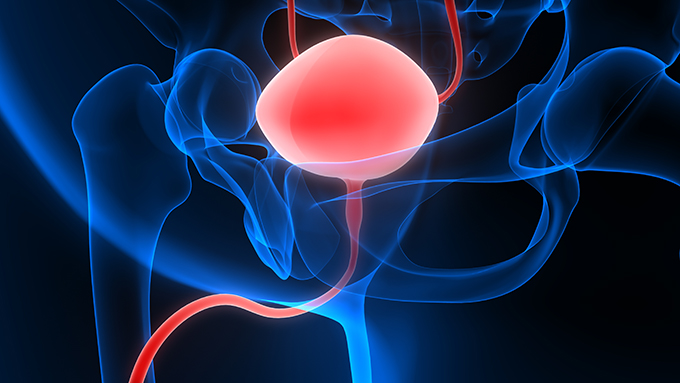
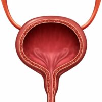
The prostate is a walnut-sized gland found beneath your bladder and in front of your rectum. It makes seminal fluid – the fluid that mixes with sperm when you ejaculate. Running directly through the prostate is the urethra, the tube that allows urine and semen to leave your body.

BPH might become aggravating, but overall, it’s not harmful

BPH becomes more common as you get older. According to UpToDate, an estimated 8% of men between the ages of 31 to 40 have an enlarged prostate. Over age 80, the rate is over 80%.

One word that should reassure you is benign. It might become aggravating, but overall, it’s not harmful. And BPH is not the same as prostate cancer. It is possible to have both an enlarged prostate and prostate cancer, but BPH on its own is very common and very treatable.

Symptoms of BPH

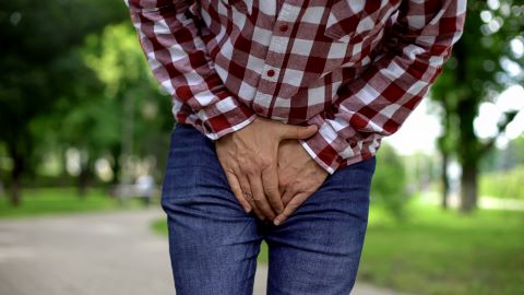
While BPH is benign, it can still have some frustrating symptoms, mostly urinary:

  • A more urgent and frequent need to urinate. That “gotta go” feeling might be stronger, and it might happen more often.
  • Frequent urination at night (nocturia). If you’re waking up every couple hours with the need to pee, that could be a sign of BPH.
  • Feeling like your bladder isn’t completely empty, even if the urine flow has stopped.
  • Straining during urination. You might feel like it takes more effort – or you have to push – in order to pee.
  • Weak urine flow or a flow that stops and starts. You might dribble or leak urine, too.
  • Bloody urine (hematuria)

BPH symptoms usually start when a man reaches his 50s. However, not all men with BPH have symptoms. Some have no problems at all. Or if they do, the symptoms don’t bother them.

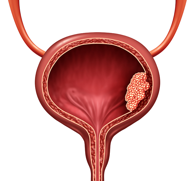
What happens when the prostate gland grows?

Basically, when the prostate gland gets bigger, it grows inward rather than outward. And this is why urination becomes a problem.

Recall that the urethra goes straight through the prostate gland. That new growth going inward can press against the urethra – squeezing it, if you will. This process narrows the path for urine flow, slowing it down or blocking it altogether.

Should you simply tolerate BPH symptoms, chalking them up to signs of aging? No. For one thing, there are a variety of treatments available, so there’s no reason to suffer with symptoms. Chances are, there’s a treatment that will work for you.

Secondly, not treating BPH can lead to other problems. Your BPH symptoms might worsen over time, eventually damaging your bladder or kidneys.

A doctor holding a clipboard and pen talks to a male patient.

Diagnosis

Doctors diagnose BPH by taking a medical history and conducting a number of tests. You’ll be asked a lot of questions, and it’s important to be honest. The tests? Some can be uncomfortable. But they’re essential for ruling out other conditions that have similar symptoms:

  • Urethral stricture
  • Prostate cancer
  • Bladder cancer
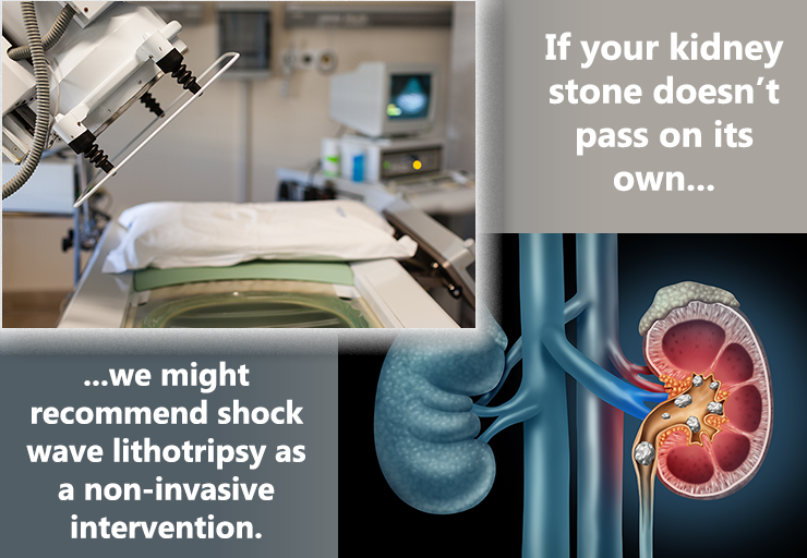
  • Kidney or bladder stones
  • Overactive bladder
  • Neurogenic bladder

Your medical history

Your doctor will want to know about your family medical history and your own experiences with urinary tract infections and prostatitis (inflammation of the prostate). They will also ask you about any over-the-counter products and prescription medications you use. You might want to make a list of these before your appointment. If you use any herbal products or nutritional supplements, make sure you mention those.

The doctor might want to observe you urinating and will likely check your abdomen to see if there are signs of an enlarged bladder.

You might also be asked to complete a questionnaire called the AUA (American Urological Association) Prostate Symptom Index or another assessment tool.

Digital Rectal Exam (DRE)

This exam can be awkward. But it only takes a few minutes, and your doctor can get a lot of useful information.

During a DRE, your doctor will place a lubricated, gloved finger into your rectum. In this way, they doctor can actually feel your prostate gland and assess its size and its texture (lumps, hard spots, soft spots).

A DRE shouldn’t hurt, but you might feel a little uncomfortable. Take some deep breaths, knowing that it will be over soon.

Lab tests

  • Urine test (urinalysis). This test is straightforward: You pee into a cup. Your urine sample is then analyzed to see if there is anything unusual.
  • Blood tests. In your blood work, your doctor will check your PSA levels. PSA stands for prostate specific antigen. Patients with benign prostatic hyperplasia (BPH) or prostatitis may have larger amounts of PSA.

Other tests

  • Urodynamic tests measure the volume and pressure of urine in the bladder and evaluate the flow of urine.
  • Uroflowmetry tells the doctor how quickly – and to what extent – you can empty your bladder.
  • A pressure flow study can help your doctor determine how much blockage there is.
  • A post-void residual (PVR) test measures how much urine stays in your bladder after urination.
  • Ultrasonography creates images of your urinary tract to show any abnormalities. Your doctor can estimate the size of your prostate this way, too.

How is BPH treated?

If it turns out you have BPH, you have lots of treatment options. We’ll run through them here, but your doctor will help you decide which one is right for you.

Watchful Waiting

This is a “wait and see” approach. If you’re just starting to have symptoms, or if they’re not bothering you that much, you might not begin treatment right away. Instead, you and your doctor will keep an eye on things. If your symptoms get worse, you can look into other treatments. If they improve (and for some men they do), that’s even better.

While you’re waiting, you can take these steps to get some relief from urinary symptoms:

  • Watch your fluid intake. Skip that glass of water at bedtime or if you’re not sure where your next bathroom will be.
  • Caffeinated drinks (like coffee, tea, and soda) and alcohol can make you have to “go.” Cut back on these.
  • Try to empty your bladder before you going to bed or heading out. If you feel like it’s not completely empty, try again a few minutes later.
  • Avoid certain medications, like tranquilizers and over-the-counter cold remedies containing decongestants and antihistamines. These can worsen urinary symptoms.
  • Avoid spicy or salty foods.
  • Watch your weight and eat a healthy diet.

Medications

Generally, two types of medications are used to treat BPH:

Alpha blockers

If your doctor recommends medication, you’ll probably start with alpha blockers. These drugs open up your urine flow by relaxing smooth muscle tissue, taking some pressure off the urethra.

Examples include terazosin (Hytrin), doxazosin (Cardura), tamsulosin (Flomax), alfuzosin (Uroxatral), and silodosin (Rapaflo).

Alpha blockers can have some side effects, such as headache, dizziness, low blood pressure, fatigue, weakness, and difficulty breathing.

Note: Erectile dysfunction (ED) drugs called phosphodiesterase type 5 (PDE5) inhibitors might interact with alpha blockers. Always check with your doctor if you’re thinking about taking these drugs together.

Alpha-reductase inhibitors

These drugs work by stopping prostate growth. It can take up to 6 months to see how well they work.

Examples include finasteride (Proscar) and dutasteride (Avodart).

Possible side effects are diminished libido, erection or ejaculation problems, depression, breast tenderness or enlargement, and reduced sperm count.

Note: If your partner is pregnant, she should never handle these drugs. Exposure can cause serious side effects for a developing fetus.

Combination Treatments

Some men, especially those with severe cases of BPH or very large prostates, take both alpha blockers and alpha-reductase inhibitors.

What about herbal treatments like saw palmetto?

You might have heard about herbal treatments, particularly one called saw palmetto. However, these aren’t usually recommended. Keep in mind that herbal treatments aren’t regulated by official agencies, like the FDA. And they can interfere with other medications you’re taking. If you’re thinking about any herbal remedies, always check with your doctor first!

Minimally Invasive and Surgical Treatments

If medications aren’t right for you, you doctor might recommend a minimally invasive treatment or surgery. Some of these procedures can be done right in your doctor’s office. Some require a hospital stay. Some men have temporary side effects, like erectile dysfunction (ED). Some men don’t. Your doctor will go over your options. Usually, treatment depends on the size of your prostate, the severity of your symptoms, and your overall health.

Keep in mind that the results of some treatment last longer than others. Ask your doctor if you might need another treatment down the road.

Prostatic Urethral Lift (PUL)

Some doctors call this technique Urolift®. With this procedure, you’ll have either local or general anesthesia. Your surgeon will then use a special needle to position special implants to hold prostate tissue back, away from your urethra. Most men go home the same day as a PUL procedure.

Transurethral Treatments

The word transurethral means “through the urethra.” In other words, excess prostate tissue is removed through your urethra. You might wonder how an enlarged prostate might be accessed through a tiny tube, but doctors use special equipment designed to fit. You’ll be given anesthesia, so you won’t feel a thing.

Transurethral Resection of the Prostate (TURP)

TURP is a common procedure. The AUA estimates that about 150,000 men in the United States undergo TURP every year.

For TURP, you’ll receive general anesthesia. During the procedure, an instrument called a resectoscope is inserted into your urethra. This device has an electrical loop at the end, which removes obstructing prostate tissue and seals blood vessels. An irrigating fluid is used to flush out the debris. Any remaining debris will pass through your urine.

You’ll probably be in the hospital for about 3 days, and you’ll have a catheter to drain urine. During your recovery, you might have some initial discomfort, feel an urgent need to urinate, or have trouble controlling urination. Usually, these symptoms clear up before long.

Some men have sexual problems after TURP, and it might take a year for your sex life to get back to the way it was before the procedure. The most common issue is retrograde ejaculation, sometimes called “dry orgasm” or “dry climax.” You’ll still ejaculate and feel pleasure from orgasm. But semen will travel backward into your bladder instead of forward out of your penis. It eventually passes with your urine. Retrograde ejaculation is harmless, but if you wish to father a child, talk to your doctor first.

Transurethral Incision of the Prostate (TUIP)

If your prostate isn’t greatly enlarged, you might have a TUIP procedure.
Your surgeon will use a laser beam or electrical current to make small cuts in the bladder neck where the urethra joins the bladder, extending into the prostate. This reduces pressure on the urethra.

You might be in the hospital for about 3 days for TUIP, and you’ll probably have a catheter for a few days, too.

Urinary tract infections, retrograde ejaculation, and ED are possible side effects of TUIP, however most issues are temporary. Additional treatment might be necessary in a few years.

(Note: When a laser beam is used, TUIP might be called TULIP (transurethral ultrasound-guided laser incision of the prostate).

Transurethral Electroevaporation of the Prostate (TUVP)

With TUVP, a tool called a resectoscope is inserted into your urethra (after you receive anesthesia). This tool emits an electrical current to destroy prostate tissue. You’ll be in the hospital for one night.

Transurethral Water-Jet Ablation (TWJA)

As the name suggests, TWJA uses strong water jets to destroy extra tissue. It’s a fairly new procedure, and you’ll have general anesthesia. Your hospital stay will likely be just one night, and you’ll need a catheter for about two days. This approach is thought to have fewer sexual side effects.

Transurethral Microwave Thermotherapy (TUMT)

With TUMT, microwaves are used to heat prostate tissue and destroy it. (Don’t worry! There’s a cooling system in place that will protect your urinary tract.) You won’t need anesthesia for TUMT, and you’ll probably go home the same day.

Ablation of the Prostate

Ablation, in BPH terms, means “destroy” excess prostate tissue using electrical, heat, or laser energy. These procedures tend to have less bleeding, and could be more suitable for men with other health issues. Here are some examples:

  • Plasma vaporization (“button procedure”). Similar to TURP, but instead of an electrical loop at the end of the device, there are two electrodes.
  • Holmium laser enucleation of the prostate (HoLEP). This procedure is also similar to TURP, but uses a holmium laser. You might be in the hospital for a night. And you might experience incontinence afterward, but this usually gets better within 6 weeks.
  • Thulium laser enucleation of the prostate (ThuLEP). ThuLEP is similar to HoLEP described above, except a thulium laser is used.
  • Holmium laser ablation of the prostate (HoLAP). This procedure is also similar to HoLEP, but the laser vaporizes excess tissue.
  • Photoselective Vaporization of the Prostate (PVP). PVP uses a special high-energy laser to vaporize excess prostate tissue. The laser is delivered through an endoscope that’s inserted into your urethra.
    The procedure is performed on an outpatient basis, and you’ll likely go home within a few hours. You should still avoid strenuous exercise for 2 weeks afterward, though.
  • Convective water vapor ablation (CWVA). This technique uses the energy in water vapor (steam) to destroy excess prostate tissue. It’s a same-day procedure that can be done at your doctor’s office, though you might need to use a catheter for a few days afterward. Many men don’t have side effects.

Surgery

Simple prostatectomy

Men who have particularly large prostates or bladder damage may undergo simple prostatectomy – the surgical removal of prostate tissue. Unlike transurethral procedures, prostate tissue is removed through an incision. This incision might be in your lower abdomen (a suprapubic or retropubic prostatectomy). Another option for the location of the incision is your perineum (the area between your rectum and scrotum). This type is called a perineal prostatectomy.

Note: Simple prostatectomy is different from radical prostatectomy

(Note: Simple prostatectomy is different from radical prostatectomy, a procedure used to treat prostate cancer. With radical prostatectomy, the entire prostate gland is removed. Simple prostatectomy just removes a portion of it.)

Nowadays, some prostatectomies are robot-assisted. However, this doesn’t mean that a robot is actually doing your surgery. Your surgeon is still in charge! However your surgeon guides the robot with a computer and follows the action on a monitor. This allows the surgeon to control the robot’s movements with great precision.

You’ll be in the hospital for a few days after an open prostatectomy. Once you’re home, you’ll need to take it easy and avoid any strenuous activity or lifting for a few additional weeks. Depending on the prostate size, many centers with robot-assisted laparoscopic techniques can now perform the surgery using the robot. The advantage of the latter is a shorter hospital stay and earlier recovery.

Some men have incontinence (urine leakage) or erection difficulties after prostatectomy. Your doctor can teach you how to do special exercises called Kegels to strengthen your pelvic floor muscles. Your doctor might also prescribe treatment for erectile dysfunction (ED).


Bladder Cancer

Bladder cancer is the 6th most common cancer in the United States, according to the Urology Care Foundation. More men than women get bladder cancer, and while it can happen at any age, it’s more common in people age 75 and older.

The most frequently diagnosed type of bladder cancer in the United States and Europe is called urothelial cell carcinoma or transitional cell carcinoma. That’s the type we’re going to talk about today.

3D Illustration Concept of Human Urinary System Bladder Anatomy

Your bladder is like a storage tank for urine. Whenever you urinate, you empty your bladder. Then it gradually fills until it’s time to urinate again. The bladder’s flexible wall is made up of five layers. The mucosa (urothelium) is the innermost layer. Behind that is the lamina propria. Next, you’ll find the muscle. Beyond that are a layer of fatty tissue and the outside layer – the peritoneum – which surrounds and protects the whole bladder.

In general, bladder cancer is classified in one of two ways, depending on whether cancer cells have reached the muscle layer. The deeper cancer grows into the wall, the more advanced it is. This classification helps us choose a path for treatment.

Non-muscle invasive bladder cancer (NMIBC)

Human bladder cancer as a urinary anatomical organ disease and malignant cells concept as a 3D illustration cutaway of body anatomy.

Most cases of bladder cancer (70% – 75%) fall into this category. In this situation, cancer cells may have penetrated the bladder’s two inner layers. But they haven’t yet reached the muscle.

Non-muscle invasive bladder cancer is sometimes called superficial bladder cancer because it is affecting only the “surface” inner layers of the bladder. However, this type of cancer can still progress to the deeper layers.

You might also see the terms T0 or T1 to describe this type of cancer.

Muscle-invasive bladder cancer (MIBC)

With this type of bladder cancer, cells have penetrated the muscle layer and, in some cases, the fat layer beyond it. Muscle invasive bladder cancer is more serious and often requires surgical bladder removal as part of treatment. You might see the terms T2, T3, and T4 associated with this type.

Regardless of the type, bladder cancer has similar risk factors, symptoms, and diagnostic procedures.

Bladder Cancer Risk Factors

What increases your risk for bladder cancer?

  • Smoking. People who smoke cigarettes are 2 to 4 times more likely to develop bladder cancer than nonsmokers. In fact, half of all bladder tumors are thought to be caused by smoking tobacco. And the more you smoke, the higher your chances of developing bladder cancer become. Even exposure to secondhand smoke raises bladder cancer risk.
  • Chemical exposure. Do you work with certain chemicals to make paint, plastics, leather, rubber, or textiles? Or are levels of certain chemicals elevated where you live? Exposure to some of them, such as dyes known as “azo” compounds, could increase your bladder cancer risk.
  • Some occupations seem to be at higher risk for bladder cancer, including hairdressers, machinists, printers, painters, and truck drivers.
  • Genetics. Bladder cancer can run in families. If you have a relative who has had bladder cancer, you might be at higher risk yourself.
  • Treatment for other cancers. If you’ve had radiation to the pelvis or taken drugs like cyclophosphamide to treat lymphoma or leukemia, you could have a higher risk for bladder cancer.

Bladder Cancer Symptoms

People with bladder cancer don’t always have symptoms. Those who do might have symptoms that mimic those of urinary tract infections, kidney stones, or other urological issues. You might have symptoms from time to time, or they might be constant. However, if you ever have any of the following symptoms, give us a call so we can do a full checkup:

  • Blood in your urine. The medical term for this symptom is hematuria, and it’s the most common symptom of bladder cancer. If you have blood in your urine, it might be a pinkish or reddish color. But sometimes, the blood can’t be seen with the naked eye. This is called microscopic hematuria or microhematuria, which is found during a urinalysis, a test of your urine that’s commonly done during an annual physical.
  • Changes in urination. You might leak urine, need to urinate more often, or feel a more urgent need to “go.” You might also have pain during urination (dysuria).
  • Pain. You might have pain in your side, your back, or your pelvic area.

Symptoms of more advanced bladder cancer include lack of appetite, weight loss, and fatigue.

Bladder Cancer Diagnosis

To diagnose bladder cancer, we start with a complete physical exam. We’ll also ask you questions about your medical history. From there, you might have one or more of the following tests:

Urinalysis. When we analyze your urine, we check for certain types of cells, like red blood cells and white blood cells. These cells can give us clues to your urologic health.

Urine cytology. We’ll use a microscope to check your urine sample for any abnormal cells.

Blood tests. We’ll do a comprehensive metabolic panel (CMP) and check your kidney and liver function.

Imaging tests. We might order a CT scan (computed tomography), MRI (magnetic resonance imaging), or kidney ultrasound to get a better picture of your, chest, pelvis, bladder, and surrounding organs.

Cystoscopy (cystourethroscopy). For screening purposes, a cystoscopy is done in our office with a flexible instrument called a cystoscope, which has a light and camera at the end. The cystoscope is placed into your bladder through your urethra. This allows us to see the inside of your bladder. We can also use the cystoscope to take a tissue sample (biopsy), which can be sent to a lab to check for cancer cells. You’ll have local anesthesia before this procedure.

We might also conduct a retrograde pyelogram, an x-ray imaging test, during your cystoscopy to get another look at your bladder, ureters, and kidneys.

Sometimes, a more rigid cystoscope is used. This type of instrument is bigger so that surgical instruments can be sent through it. If we use a rigid cystoscope, you’ll have full/general anesthesia.

Transurethral resection of bladder tumor (TURBT)

A TURBT procedure is similar to the cystoscopy described above, which allows us to examine your urethra and bladder. We go ahead with TURBT if previous testing suggests that you have cancer. TURBT provides us with more information about your cancer, such as tumor size, number, and location, so we can make further treatment decisions. Tumors can also be removed during TURBT.

We perform TURBT procedures at a hospital, and you’ll have general or spinal anesthesia beforehand. You’ll probably be able to go home the same day. You might have a catheter to drain urine for a day or two. We’ll show you exactly how to manage it.

Blue light cystoscopy. This test also uses a cystoscope. First, the doctor places a special imaging solution into your bladder. After an hour or so, the doctor examines your bladder with the cystoscope, but uses a blue light. The solution makes cancer cells easier to see in blue light.

Bladder Cancer Treatment

As we mentioned above, your bladder cancer treatment will largely depend on how deeply cancer cells have penetrated your bladder wall. We’ll also consider your overall health, and the type of tumor you have.

Before any treatment begins, we’ll talk with you about potential complications and the impact treatment may have on your quality of life.

Elderly husband giving to his sick wife with glass of water pills at home

Treatment of Non-Muscle-Invasive (Superficial) Bladder Cancer (NMIBC)

To treat non-muscle invasive bladder cancer, we’ll start by removing as much of the tumor as we can.

Transurethral resection of bladder tumor (TURBT)

You will likely have a TURBT during your screening process (see above). TURBT for treatment is similar. You’ll be given anesthesia and we’ll pass a rigid cystoscope (the larger kind that doesn’t bend) through your urethra to reach the bladder. Since we can pass surgical instruments through the cystoscope, we won’t have to make any incisions. We might also conduct a blue light cystoscopy at this time for further evaluation.

During the procedure, as much of the tumor (or tumors, if you have more than one) will be removed as possible. Areas that look suspicious for cancer can also be removed.

As with a diagnostic TURBT, you might use a catheter while your bladder heals.

A few weeks after this TURBT procedure, we might recommend another one. We do this to make sure all of the tumor is gone. If any cancerous tissue has been missed, we can remove it.

Adjuvant bladder cancer therapy

Once we’ve removed the tumor(s), you’ll likely have additional treatment called adjuvant therapy.

Why? Bladder cancer recurrence is fairly common. In fact, about half of people with bladder cancer see their cancer return within a year. Adjuvant therapy can reduce the risk of recurrence.

Intravesical Therapy

With intravesical therapy, cancer-fighting drugs are placed directly into your bladder through a catheter instead of in your bloodstream with an IV line. They stay in your bladder for an hour or two, and then they are drained out. The goal is to attack any remaining cancer cells that might have broken away from the tumor and stop them from starting new tumors in the bladder wall.

Intravesical therapy may be accomplished through immunotherapy or chemotherapy. The type and duration of treatment you receive will depend on your personal situation and whether your cancer is considered low-, intermediate-, or high-risk.

Immunotherapy for Bladder Cancer

Intravesical immunotherapy taps the power of your own immune system to fight cancer cells using a drug called Bacillus Calmette-Guerin (BCG). Like chemotherapy drugs, BCG is directed into your bladder through a catheter.

You might need several courses of immunotherapy, and overall treatment could last about 6 weeks. We might also repeat it periodically for a few years as a maintenance treatment. Side effects include urinating more frequently, pain during urination, joint pain, and flu-like symptoms. Usually, these side effects go away within 48 hours.

Chemotherapy for Bladder Cancer

Intravesical chemotherapy (“chemo”) usually involves the use of one of the following drugs: mitomycin C or gemcitabine. These drugs affect only the bladder lining.

The first course of intravesical chemotherapy is generally done within 24 hours of TURBT. If your risk of recurrence is low, you might have intravesical chemotherapy just once. But if your risk is higher, we might repeat the process once a week for about 6 weeks. We might also repeat it again in 1 to 3 years as maintenance therapy.

Side effects of chemo include more frequent urination, painful urination, flu-like symptoms, and skin rash. Usually, these side effects are temporary. If you develop a skin rash that becomes severe, we can prescribe cortisone therapy or change the type of chemotherapy drug we use.

Treatment of Muscle-Invasive Bladder Cancer (MIBC)

Because cancer cells have penetrated the muscle layer, muscle-invasive bladder cancer is a more advanced stage of the disease. Your treatment will depend on how much the cancer has progressed and your overall health. But typically, treatment for this type of cancer includes chemotherapy, surgery to remove the bladder, and, in some cases, radiation therapy.

Chemotherapy for Muscle-Invasive Bladder Cancer

Unlike chemotherapy for non-muscle-invasive bladder cancer, chemotherapy drugs for MIBC are directed right into your bloodstream intravenously. Cisplatin is the most commonly used drug, although it might be combined with another drug.

Usually, you can receive chemotherapy in an outpatient setting. You might need a few cycles of chemo, and we’ll let you know what you can expect.

Chemotherapy can have side effects. You might feel fatigued and weak, and you might not feel like eating much. Nausea, diarrhea, and hair loss are common. You might also get sores on your mouth or lips, and you’ll be at higher risk for infections, bruising, and bleeding. We’ll be monitoring all these symptoms with you, and we’ll be here to help you manage them.

The time frame for chemotherapy depends on your other treatments.

  • Neoadjuvant cisplatin-based chemotherapy (NAC). Neoadjuvant chemo is given to shrink your tumor and eliminate any random cancer cells as much as possible before bladder surgery. Generally, surgery is scheduled within 12 weeks after you finish chemo.
  • Adjuvant chemotherapy. Not everyone can have chemotherapy before their surgery. In these cases, you might have adjuvant chemotherapy, which occurs after surgery. Similar to neoadjuvant therapy, the chemo drug cisplatin is used in adjuvant chemotherapy.

Radical Cystectomy (Surgical Removal of the Bladder)

It’s common for patients with MIBC to have their bladder surgically removed, either partially or completely. If cancer has spread outside the bladder, pelvic organs and/or tissues that are involved might need to be removed as well, such as lymph nodes, part of the urethra (the tube that allows urine to exit the body). Other areas that may be involved are the prostate and seminal vesicles in men, and reproductive organs adjacent to the bladder in women.

Radical cystectomy may be performed in one of two ways:

  • Open cystectomy. An incision is made in your abdomen and your bladder is removed through that opening.
  • Robotic cystectomy. During a robotic procedure, smaller incisions are made, and a robot will hold the surgical instruments. The robot is controlled by the surgeon at all times. People who have robotic surgeries tend to have less pain and lose less blood.

After surgery, you’ll need to take it easy and give yourself time to heal. You might be in the hospital for about a week and then spend the rest of your recovery period at home. We’ll give you complete instructions for your recovery and prescribe medicine for pain. Most patients can go back to their day-to-day lives within 6 weeks of surgery.

Urinary Diversion

When you have your bladder removed, your surgeon will create a way for urine to leave your body. This process is called urinary diversion. Depending on your situation and preferences, this can be accomplished in a few ways:

  • Ileal conduit/urostomy. With this method, the surgeon uses a piece of your bowel (intestine) to make a stoma (an opening) on your skin near your stomach. Your ureters, which typically connect the kidneys to the bladder, will be diverted to the stoma instead. Urine will drain into a special collection bag that you will wear outside your body and empty throughout the day.
  • Continent cutaneous reservoir. Your surgeon will create a pouch from a piece of your intestine and attach it to a stoma in your abdomen. You’ll use a catheter to drain your urine.
  • Orthotopic neobladder. In this case, your surgeon will make a neobladder – an internal pouch – and connect your ureters to it. With a neobladder, you will be able to urinate like you did before. There is no collection bag or catheter involved.

Before you leave the hospital, you’ll receive full instructions on how to manage and care for your urinary diversion method.

Partial Bladder Removal

In fewer than 5% of cases, patients have a partial cystectomy, which means that only part of the bladder is removed. This procedure may be possible for people whose tumor is in one concentrated area.

After a partial cystectomy, you should be able to urinate the same way you used to.

Bladder Preservation

There are occasionally cases of MIBC when the bladder can be spared due to the limited spread of cancer into the muscle wall.

If you’re undergoing bladder preservation, you’ll have a TURBT procedure similar to what we’ve described above (see the section on non-muscle-invasive bladder cancer). However, the surgeon will cut tumors deeper into your bladder wall. You’ll also have chemotherapy – as described above – and in addition, radiation therapy. Radiation therapy is carried out over the course of several weeks. It doesn’t hurt, but sometimes causes nausea or fatigue afterward.

Bladder preservation has a higher risk of cancer recurrence. The American Urological Association estimates that 30% of patients who follow this treatment path have a recurrence of MIBC.. We will monitor your progress with routine CT scans, cystoscopy, and urine cytology.

We work with our patients to develop a treatment plan that we feel will provide the best quality of life and the lowest risk of recurrence.

Sexual Function After Cystectomy

In both men and women, an intricate network of nerves needed for sexual function are located close to the bladder. When surgery requires disrupting the nerves, it can lead to erectile dysfunction (ED) in men and vaginal dryness in women.

A surgeon’s goal is to keep these nerves intact. However, if this is not possible – or if you have any sexual difficulties after surgery – such issues can be treated. For example, many men can take medications to restore their erections. And women may use a lubricant to make intercourse more comfortable. Don’t hesitate to ask us about nerve-sparing procedures or any sexual issues that occur after surgery.

After Treatment

No matter what type of bladder cancer treatment patients have, they need to be monitored closely with regular follow-up appointments and tests. It’s important that we monitor patients closely in order to catch a recurrence as soon as possible.

Most likely, we’ll conduct imaging, cystoscopy, lab, and urine cytology tests every 3 to 6 months for up to 4 years. If there is no cancer recurrence, then we’ll see you annually after that.

Keep in mind that we are always here if you have questions or need additional help. For example, we can guide you on making good lifestyle choices to keep you healthy. And we can put you in touch with cancer support groups and counselors, which can help you cope with the day-to-day and the emotional aspects of cancer.

Resources

American Urological Association

Chang, S.S., et al.
“Diagnosis and Treatment of Non-Muscle Invasive Bladder Cancer: AUA/SUO Joint Guideline (2020)”
(Published: 2016. Amended: 2020)
https://www.auanet.org/guidelines/guidelines/bladder-cancer-non-muscle-invasive-guideline

Chang, S.S., et al.
“Treatment of Non-Metastatic Muscle-Invasive Bladder Cancer: AUA/ASCO/ASTRO/SUO Guideline (Amended 2020)”
(Published: 2016. Amended 2020)
https://www.auanet.org/guidelines/guidelines/bladder-cancer-non-metastatic-muscle-invasive-guideline

Medical News Today

Fletcher, Jenna
“What to expect with bladder removal surgery”
(May 31, 2018)
https://www.medicalnewstoday.com/articles/321994

National Institute of Diabetes and Digestive and Kidney Diseases

“Urinary Diversion”
(Last reviewed: June 2020)
https://www.niddk.nih.gov/health-information/urologic-diseases/urinary-diversion

UpToDate.com

Black, Peter, MD, FACS, FRCSC and Wassim Kassouf, MD, CM, FRCS
“Patient education: Bladder cancer treatment; invasive cancer (Beyond the Basics)”
(Topic last updated: May 19, 2020)
https://www.uptodate.com/contents/bladder-cancer-treatment-invasive-cancer-beyond-the-basics

Kassouf, Wassim, MD, CM, FRCS and Peter Black, MD, FACS, FRCSC
“Patient education: Bladder cancer treatment; non-muscle invasive (superficial) cancer (Beyond the Basics)”
(Topic last updated: April 1, 2020)
https://www.uptodate.com/contents/bladder-cancer-treatment-non-muscle-invasive-superficial-cancer-beyond-the-basics

Lotan, Yair, MD and Toni K Choueiri, MD
“Patient education: Bladder cancer diagnosis and staging (Beyond the Basics)”
(Topic last updated: April 9, 2019)
https://www.uptodate.com/contents/bladder-cancer-diagnosis-and-staging-beyond-the-basics

Urology Care Foundation

“Immunotherapy and Bladder Cancer”
https://www.urologyhealth.org/resources/immunotherapy-and-bladder-cancer

“What is Muscle Invasive Bladder Cancer (MIBC)?”
https://urologyhealth.org/urology-a-z/m/muscle-invasive-bladder-cancer

”What is Non-Muscle Invasive Bladder Cancer?”
(Updated: August 8, 2020)
https://urologyhealth.org/urology-a-z/n/non-muscle-invasive-bladder-cancer


Erectile Dysfunction

Erectile dysfunction (ED) – you know the term. It’s when a man can’t get – or keep – an erection hard enough for satisfying sex. Your father or grandfather might have called it impotence, if they talked about it at all.

A man sits on the edge of a bed with his hands cradling his face. A woman sits under the covers in the background.

If you can’t get an erection at all, no matter what, that’s ED. But a partial erection – one that’s not as firm as you’d like it to be – is ED too. Maybe it only happens once in a while. Or maybe it only happens in certain situations – like when you’re with someone but not when you masturbate. It’s all ED.

Understandably, men are typically concerned about ED. Your sex life is likely to suffer – or at least change quite a bit. Your sense of masculinity may be bruised. You may be worried about satisfying your partner. And what about your overall physical health?

Don’t panic! ED is actually quite common. Most men experience it at some point in their lives. And often, it’s very treatable.

The American Urological Association estimates that 30 million men have erectile dysfunction

How common is ED?

It’s the most common sex problem men discuss with their doctors. The American Urological Association (AUA) estimates that 30 million men have ED.

ED is more common if you’re older, but younger men get it, too. If you’re not as healthy as you could be, the odds you’ll be dealing with ED go up. If you have diabetes, heart disease, or kidney disease, you’re at higher risk. Psychological issues, like depression and anxiety, can play a role as well.

Why can’t I get (or keep) a strong erection?

Ideally, here’s what happens to your penis when you get aroused: smooth muscle tissue relaxes and arteries widen. Additional blood starts to flow in. This extra blood makes your erection firm. Veins temporarily close, keeping the blood inside the penis until you ejaculate or the stimulation stops. Then the veins open up again and the blood flows back into your body.

ED is often a blood flow problem. The smooth muscle tissue might not relax or the arteries don’t open enough. Or, arteries might be blocked by plaques, as is the case the atherosclerosis (hardening of the arteries), making it more difficult for blood to flow in. In some cases, veins don’t constrict enough, so blood can’t stay in the penis.

What causes ED?

There are a number of possibilities here:

Vascular disease

Vascular disease involves damage to your circulatory system – the arteries and veins which carry blood throughout your body. Remember, “hard” arteries are “clogged” with plaques that form on your artery walls. Vascular disease makes it harder for blood to flow through your blood vessels. Less blood flow typically means a softer erection.

This is a common way for vascular disease to lead to ED. Your risk for vascular disease increases as you get older. An estimated 50 to 60 percent of men over 60 have ED due to vascular disease.

You’re more likely to face vascular disease if you have:

  • Diabetes
  • High blood pressure (hypertension)
  • High cholesterol
  • A history of smoking

Diabetes

When your blood sugar is too high, it can damage nerves and blood vessels throughout your body, including those needed for a firm erection. Your penis might not “get the message” from your brain to start an erection. And if an erection does get triggered, you might not have sufficient blood flow to keep it going. Diabetes is a huge ED risk factor. About 60 percent of men with diabetes have trouble with erections.

Drugs

Lots of medications men take have sexual side effects, which can include ED. If you take blood pressure drugs, heart medications, antidepressants, tranquilizers, or sedatives, you might see some changes in your erections. Over-the-counter meds that you buy at the pharmacy can have similar effects.

If you drink alcohol or use illicit drugs, you should watch your intake. These can cause ED, too.

Kidney and liver disease can affect your hormones and your sex life

Hormone imbalances

Most men know testosterone affects sexuality. It may surprise you though that it’s pretty rare for low testosterone to cause ED or low sex drive. Still, it does happen. Check with your doctor.

Prolactin is another hormone to watch. Men with pituitary gland tumors may have too much prolactin, which in turn reduces testosterone levels.

Kidney and liver disease can also affect your hormones and your sex life.

Obesity

We all know obesity is a growing problem in the United States. Maybe you’re struggling with your weight. You’re not alone. In 2017-2018, the Centers for Disease Control and Prevention (CDC) reported that 42% of Americans were obese – in other words, they had a body mass index (BMI) of over 30. Carrying extra weight puts a lot of stress on your body. The result can be heart disease, diabetes, and yes, ED.

Neurologic conditions

Some neurologic conditions, like paraplegia and stroke, interrupt nerve impulses from the brain to the penis. In other words, the penis won’t “know” that there’s a reason to become erect. Other neurologic conditions, such as multiple sclerosis (MS), Parkinson’s disease, and Alzheimer’s disease can lead to ED, too.

Pelvic trauma, surgery, radiation therapy

If you’ve had pelvic surgery, radiation treatment for cancer, or trauma to the pelvic region or spinal cord, you might experience ED.

For example, if you have prostate cancer, you might have your prostate removed surgically. Unfortunately, the prostate is surrounded by nerves needed for erections. Surgeons do their best to keep as many nerves intact as they can (a technique called nerve-sparing). But many men still have some degree of ED afterward. The situation usually improves, but it takes time – sometimes up to 18 months.

The situation is similar if you have a radical cystectomy for bladder cancer.

Peyronie’s disease

Men with Peyronie’s disease have a noticeable curve to their erect penis due to areas of hardened scar tissue (plaques) that form just beneath the skin’s surface. Some men with Peyronie’s develop painful erections or ED.

Venous leak

Good blood flow into the penis is critical for a firm erection. But that’s only half of the story. The blood needs to stay in place to keep a strong erection. Sometimes, there’s a problem with the veins, and they can’t keep the blood in. So it leaks back into your body. The result? A softer erection, or no erection at all.

Psychological and emotional issues

Stress and anxiety can have a huge impact on our health, including our sex lives. If you’re worried about your relationship, your kids, your job, or just life in general, that might transfer to the bedroom.

Add in anxiety about ED and things can go downhill quickly. If you’re in bed with your partner and worried about your performance, it’s going to be tough to relax – and that’s a recipe for ED.

A doctor holding a pen and clipboard talks to a male patient.

Diagnosing ED

We know. It’s tough for many men to talk with a doctor about the possibility of ED. But your doctor has heard it all. And talking with your doctor is the first step in doing something about the issue.

Your doctor will ask the usual questions about your medical history and your lifestyle. Do you smoke? How much exercise do you get? What’s your diet like? Your doctor will also need some details about your erections. How often do you have trouble getting or staying firm? Can you get an erection in some situations (like when you masturbate), but not others? How has your sex drive been? Do you have any pain?

Again, your doctor has heard it all. Men are treated for ED every day. You want your treatment to be as effective as possible, so answer the questions with as much honesty and detail as you can muster.

Lab tests will probably be part of this too. You’ll likely have blood and urine tests. They’ll want to check how your liver, kidneys, and thyroid are working. You might even have an ultrasound to check your blood flow.

How Is ED Treated?

We’ve all seen the ads for ED drugs. But pills are only one way to treat ED. Treatments can even be combined. Work with your doctor to find the best approach. Again, the more information you provide and the more questions you ask, the more effective your treatment is likely to be.

Lifestyle changes and natural treatments

Sometimes, ED can be managed by simple lifestyle changes. For example, if you have diabetes, then controlling your blood sugar may improve your erections. Or, if you’re taking a medication with sexual side effects, tweaking your prescription might help. (Never make medication changes on your own. Always check with your doctor.)

These basic changes are the first treatment options for many men:

Close up of a man's face lying on a pillow. His lover's mouth hovers close above him.

  • Quit smoking. Your doctor can help tailor a smoking cessation plan that is right for you. You’ll feel better. You’ll look better. Your wallet will thank you.
  • If you use recreational drugs, stop. Your doctor can help you here, too.
  • Do you drink a lot? Does someone else in your life think you should cut back? Give it a try. Or stop completely. Again, your doctor can help you.
  • Exercise! It’s the closest thing there is to a wonder drug. Start exercising (or increase your current effort even modestly) and you’ll almost immediately lower your risk for health problems. Chances are you’ll feel better too. You might lose a few pounds. Maybe your pants will fit a little better. Exercise doesn’t have to be a grind to be effective. Play some basketball. Go for a bike ride. Walking is excellent exercise.
  • Change your diet. Think salads, fruits, and veggies. Whole grain breads and pastas. Soy, beans, seeds, nuts, olive oil, and cold-water fish (like salmon, tuna, sardines, halibut, and mackerel). Avoid fast food, processed foods, caffeine, and sugar.You don’t have to make drastic changes all at once. Try a salad for lunch twice a week instead of drive through. Have an apple or a peach for dessert instead of that chocolate-chip cookie. Pour yourself a glass of flavored seltzer water instead of soda or fruit punch. Get a cookbook full of healthy recipes and experiment with foods you’ve never tried before. (This can be especially fun with your partner or friends.)

Sex therapy and counseling

If anxiety, depression, or another mental health issue is triggering your ED, your doctor might suggest counseling or sex therapy. Therapy can give you a chance to talk things over with a professional and learn some coping strategies. If you have a partner, you might bring them with you. Together, you can work on your communication and your relationship.

A physician works on papers. Focus is on a plaque in the foreground with Urologist written on it.

Pills

Yes, you’ve seen and heard commercials for ED drugs. In the United States, there are four oral medications used to treat ED: sildenafil (Viagra), vardenafil (Levitra), tadalafil (Levitra), and avanafil (Stendra).

They’re called phosphodiesterase type 5 inhibitors – PDE5s inhibitors for short. And while they are all used a little differently, they work in similar ways, by relaxing smooth muscle tissue in your penis. Then, enough blood can flow in for a firmer erection.

Usually, men take PDE5 inhibitors about an hour or two before sex, but you should always follow your doctor’s instructions.

PDE5 inhibitors are effective for about 70% of men, but they’re not safe for everyone. Men who take nitrates for heart conditions should never take PDE5 inhibitors. Combining PDE5 inhibitors with nitrates can cause a dangerous drop in blood pressure. If you take nitrates, you will need to use a different ED treatment.

Pay attention to side effects of your ED medication

Are there side effects? Yes, there can be. Some men get headaches or muscle aches when they take these drugs. Others get a stuffy nose, a flushed face, or indigestion. For most men, these effects are mild.

Men who take Viagra might notice temporary vision changes, where things look like they’re in a blue-green filter. If your visual acuity (how well you see) changes, you must stop taking the medication and inform your doctor immediately. Cialis users sometimes experience back pain.

If any side effects give you trouble, reach out to your doctor and ask whether another ED treatment is right for you.

Self-injections

The medical term for this approach is intracavernosal injection (ICI). The success rate for this method is about 85%, according to the American Urological Association (AUA). As with all medications, however, the response will depend on many different factors, including the severity of the condition and the medication dose.

A woman and man sit on a couch, looking lovingly at each other. The man has his arm around the woman's shoulders.

If you take this route, you’ll use a tiny needle to inject a medication called alprostadil into the side of your penis. Sometimes, other medications are mixed with alprostadil, so you might hear terms like bimix or trimix, depending on what is used. Ask your physician about the differences between these medications and how they work.

After the injection, an erection usually starts within 15 minutes and can last up to two hours. Don’t be alarmed if your erection lasts a while after you ejaculate. That can happen.

However, if you have a rigid erection that lasts longer than 4 hours, head to your local emergency department. This condition is called priapism and you’ll want medical attention ASAP.

If you use ICI, make sure you space each injection at least 24 hours apart. Up to 3 injections a week should be safe but ask your doctor about the right plan for you. Using ICI too often can scar the penis, which can make erections more difficult later on.

The idea of giving yourself an injection in the penis might make you queasy, but lots of men manage their ED this way. Your doctor will teach you how to do it safely for effective results and minimal pain. Your partner can also be taught how to do it.

Urethral Suppositories (MUSE)

The drug alprostadil (also used in injections) can come in suppository form, too. With the MUSE intraurethral method, you place a pellet into your urethra – the tube that allows urine and semen to leave your body. An erection usually starts within 10 minutes, when the drug gets absorbed and moves from the urethra into the erection chambers, the corpora cavernosa. Typically, the erection lasts for 30 to 60 minutes.

As with self-injections, your doctor will show you how to use MUSE safely and effectively. Side effects may include pain in the penis, testicles, legs, and the area between the scrotum and rectum. Some men feel warmth or burning sensations in the urethra, and the penis might redden. There could be some minor bleeding, too, if you don’t place the pellet correctly.

Testosterone therapy

If your testosterone levels are lower than normal (below 300 ng/dL), your doctor might suggest testosterone replacement therapy (TRT).

Testosterone is an important hormone for men. It drives male-typical characteristics like a lower voice and body hair distribution. It also drives your libido.

A woman and a man sit on a couch, smiling toward the camera. The woman leans her head against the man, who has an arm around her shoulders.

But in some cases, men’s bodies don’t produce enough testosterone. Also, men’s testosterone levels naturally drop as they get older. It’s a normal part of aging.

Your doctor might start you on TRT if you have other symptoms of testosterone deficiency, like low sex drive, depressed mood, or fatigue. You’ll follow up with your doctor periodically to see how well it’s going.

Don’t get duped! You might find over-the-counter products that claim to contain testosterone to improve sexual performance. Lots of men think about trying these on their own, without seeing a doctor. But it’s essential for your doctor to measure your testosterone levels and monitor them over time. Your doctor may also want to conduct other important blood tests.

Testosterone should always be taken by prescription. Think about it. Do you really want to trust your sexual health to the convenience store on the corner or a shady website?

Vacuum erection devices (VEDs)

If you don’t do well with medications – or can’t take them – you might try a VED, a simple mechanical tool. As the name suggests, a VED triggers an erection by using a vacuum to bring blood to your penis.

The basic components of a VED are a plastic tube, a small hand pump, and a rubber O-ring. Here’s how they work together:

  • When you’re ready to have an erection, you place your penis into the plastic tube, and hold it close to your body.
  • Next, run the hand pump for about one to three minutes.
  • Once your penis is fully erect, you place the O-ring around the base of your penis to keep the blood inside.
  • From there, you can remove the tube and have intercourse.
  • When you’re done, remove the O-ring.

VEDs have their pros and cons. On the pro side, they can be used at any VEDs have their pros and cons. On the pro side, they can be used at any time, so you can always be ready for sex. You don’t need to take any medication, and there’s no surgery involved. And if used properly, about 75% of men have success, according to the AUA. Your doctor will show you how to use it. Again, for any individual patient, the response depends on the severity of the condition and many other factors that your doctor can discuss with you.

On the con side, VEDs can be a little cumbersome. They take about 5 to 10 minutes to set up, which means you and your partner will have to take a short break during lovemaking so you can get an erection started. Some couples find this interruption less than romantic, and it might take away some of the excitement and spontaneity.

Other drawbacks to the VED: Your erection might look a bit floppy because there’s no erection/additional blood stored between the O-ring and your body. Some men find that their ejaculation feels different. You could feel some coldness or numbness in your penis, too.

Note: Sometimes, a man’s body shape makes it difficult to use a VED. Also, men who have problems with blood clotting and those who use blood thinners shouldn’t use VEDs.

Revascularization surgery

Younger men with ED caused by pelvic trauma may benefit from revascularization surgery. This process involves using an artery from the abdomen to create a “bypass” in the penis.

Revascularization is a very uncommon and specialized surgery that is ideal for young men, often trauma patients, who do not have general vascular disease since the “new” connection needs to be “atherosclerosis free” to be able to bring blood to the penis.

Physicians and assistants perform a surgery.

Penile implants (prostheses)

What, exactly, is an implant? It’s a device that’s surgically implanted into your penis and helps you create an erection on demand.

Implants come in two types:

  • Malleable implants are semi-rigid bendable rods, usually made from silicone. When you want an erection, you simply move your penis into an upward position. When you’re finished with sex, you can move it back down.
  • Inflatable implants are cylinders that fill with fluid to create an erection. You control the timing by using a special pump that is also surgically implanted, usually in your scrotum. You can start an erection with just the press of a button. After sex, you can push the same button to deflate the cylinders.

Getting an implant is a big decision. Once you have one, there’s no going back. That’s because the rods or cylinders replace two chambers in the penis called the corpora cavernosa. These are the chambers that normally fill with blood during an erection. During implant surgery, the corpora cavernosa are removed completely.

Implant surgery is fairly straightforward. Chances are, you’ll spend a night in the hospital, although you might go home the same day. You’ll probably have some pain, bruising, and swelling for a few weeks. Your doctor will give you special instructions about lifting things and exercising. You’ll need to wait about 8 weeks before having sex again.

Like any surgery, implants have risks. You might develop an infection. There’s also a slim chance that the device will malfunction, and you might need surgery again to fix the problem. However, most men with implants have successful, satisfying sex lives for many years after surgery.

Implants do have high satisfaction rates among men and their partners. In fact, some partners might not even know you have an implant. And orgasm should feel the same.

How about supplements?

Some men find it helpful to take supplements and herbs, but your mileage may vary. No matter what, always check in with your doctor before taking any product. Supplements aren’t regulated by the FDA the way drugs are, and you might not know how a product will interact with other health conditions or medications.


Genitourinary Syndrome of Menopause

Genitourinary Syndrome of Menopause (GSM), formerly known as vulvovaginal atrophy (VVA) or atrophic vaginitis, is a common condition that affects many women during and after menopause. It is primarily caused by the hormonal changes that occur during menopause, specifically the decline in estrogen levels. Estrogen plays a crucial role in maintaining the health of the genital and urinary tissues in women.

jonathan borba n1b6ftpb5eg unsplash

What are the symptoms of Genitourinary Syndrome of Menopause?

GSM encompasses a range of symptoms and changes in the genital and urinary tract, including:

  1. Vaginal dryness: Reduced estrogen levels can lead to a decrease in vaginal lubrication, making sexual intercourse uncomfortable or painful.
  2. Vaginal atrophy: The vaginal walls become thinner, less elastic, and more fragile, which can cause itching, burning, and discomfort.
  3. Genital discomfort: Women with GSM may experience pain, soreness, or a sense of irritation around the vulva.
  4. Dyspareunia: Painful sexual intercourse is a common symptom of GSM due to the thinning and dryness of the vaginal tissues.
  5. Urinary symptoms: Some women with GSM may experience urinary problems, such as urinary urgency, frequency, or recurrent urinary tract infections, as the tissues in the urinary tract also become affected by the estrogen deficiency.
  6. Recurrent urinary tract infections: Changes in the urinary tract can make women more susceptible to urinary tract infections.

Treatment for Genitourinary Syndrome of Menopause

GSM treatment typically involves the use of estrogen therapy, which can be administered in various forms, including vaginal creams, tablets, rings, or patches. These treatments can help restore the thickness and elasticity of the vaginal tissues and alleviate many of the associated symptoms.

Additionally, pelvic floor muscle training, lubricants, moisturizers, selective estrogen receptor modulators, vaginal DHEA and vaginal vibratory devices may also aid in the therapy.

Is Vaginal Estrogen Safe?

The use of vaginal estrogen therapy for the treatment of Genitourinary Syndrome of Menopause (GSM) is generally considered safe and effective when used as prescribed and under the guidance of a healthcare provider. However, as with any medical treatment, there are potential risks and benefits to consider.

Here are some key points to keep in mind regarding the safety of vaginal estrogen therapy:

1. Localized Treatment: Vaginal estrogen therapy is a localized treatment, meaning it primarily affects the vaginal and urinary tissues. The estrogen is absorbed in smaller amounts compared to systemic (whole-body) hormone replacement therapy (HRT), which minimizes the risk of some systemic side effects.

2. Effectiveness: Vaginal estrogen therapy is highly effective at relieving the symptoms of GSM, such as vaginal dryness, atrophy, and discomfort, as it helps restore the health of the vaginal and urinary tissues.

3. Low Absorption: When used as directed, vaginal estrogen preparations have minimal absorption into the bloodstream. This reduces the risk of systemic side effects associated with estrogen therapy.

4. Minimal Systemic Effects: While systemic estrogen therapy (like a pill or pellets under the skin) may have potential risks like an increased risk of blood clots, breast cancer, and cardiovascular issues, these risks are eliminated with vaginal estrogen because of the lower systemic absorption.

The latest study published in 2023 looking at a large group of women with previous history of breast cancer, the use of vaginal estrogen did NOT increase their risk of cancer recurrence.1

References

  1. Agrawal P, Singh SM, Able C, Dumas K, Kohn J, Kohn TP, Clifton M. Safety of Vaginal Estrogen Therapy for Genitourinary Syndrome of Menopause in Women With a History of Breast Cancer. Obstet Gynecol. 2023 Sep 1;142(3):660-668. doi: 10.1097/AOG.0000000000005294. Epub 2023 Aug 3. PMID: 37535961.
  2. https://www.prosayla.com/articles/genitourinary-syndrome-of-menopause-gsm

Written by Dr. Vladislav Gorbatiy


Interstitial Cystitis/Bladder Pain Syndrome

What is Interstitial Cystitis?

Interstitial cystitis (IC) is sometimes called bladder pain syndrome in large part because a painful bladder is one of the most prominent symptoms. People with IC can also feel a frequent, urgent need to urinate. Some people feel mild discomfort, but others may have cases so severe that it disrupts their day-to-day lives.

IC can be a challenging condition because experts aren’t sure what causes it, and that makes it difficult to treat.

A woman lies curled up in fetal position on a bed

What Causes Interstitial Cystitis?

The exact cause of IC isn’t known, but scientists have some hypotheses. Some think IC is caused by abnormalities in the bladder lining. However, they’re not sure whether IC causes these abnormalities or if the abnormalities cause IC.

Other experts believe that nerves in the bladder itself and in areas outside the bladder (such as in the abdomen or pelvis) become more sensitive.

Scientists have also noted that, in some cases, IC develops after a urinary tract infection (UTI), vaginitis (inflammation of the vagina), or prostatitis (inflammation of the prostate). It might also start after surgery or trauma in the bladder/pelvic region.

IC can be a challenging condition because experts aren’t sure what causes it, and that makes it difficult to treat.

What are the Symptoms of Interstitial Cystitis?

The most common symptoms of IC are the following:

  • Pain. Pain or discomfort may be located in or around the bladder, abdomen, or urethra (the tube that allows urine to exit the body). The pain may be mild or severe, and it could be occasional or constant. Pain might subside when you urinate.
  • Frequent and urgent urination. Some people with IC need to urinate several times an hour. You might also feel like the need to urinate is more urgent.

For most people, IC symptoms come on gradually, becoming more intense over a period of months. However, symptoms can be severe from the start.

Symptoms can also get better or worse for a variety of reasons. For example, stress might aggravate symptoms. Or a spicy meal, coffee, or alcohol may cause greater discomfort. Some activities like having sex, exercising, or sitting for a long time could trigger symptoms. Women with IC may see symptoms flare during their menstrual periods.

IC can be especially challenging for people with other chronic pain conditions, such as irritable bowel syndrome (IBS), fibromyalgia, endometriosis, and prostatitis. Sometimes, IC symptoms flare when these other conditions flare.

Coping with IC can be difficult. It can affect daily life, relationships, and sleep patterns.

How is Interstitial Cystitis Diagnosed?

There is no definitive exam for diagnosing IC, and it can take time to rule out other conditions, like kidney stones or a bladder infection. IC can be diagnosed once a person has had symptoms for six weeks and other conditions that cause similar symptoms can be ruled out. But in general, the diagnostic process follows these steps:

IC can be diagnosed once a person has had symptoms for six weeks

Medical History

We’ll talk to you about your symptoms, their severity, their effects on your everyday life, your health history, your diet, and any medications you take. We’ll also ask you questions about urination, such as how often you have to urinate and how much urine you pass. You might be asked to fill out a questionnaire as well.

Some of these questions may sound intrusive or be somewhat embarrassing. But your complete, candid answers will help us tailor the right treatment plan for you.

Physical Exam

We’ll likely conduct pelvic and rectal exams, as gently as possible. Be sure to tell us if you’re experiencing any discomfort during these exams.

We might conduct a post-void residual urine test. This tells us how much urine stays in your bladder after you urinate. It can be done with a catheter or through an ultrasound.

A man holding his groin in discomfort

You might also undergo neurological testing. The brain and the bladder work closely together, sending messages back and forth through your central nervous system. These messages may indicate that the bladder is full, that urine needs to be held, or that urine can be released. Through neurological testing, we can find out how well your brain, bladder, and nerves are communicating.

Other Urological Tests

We might conduct additional tests to rule out other health conditions. Here are some examples:

  • A urine test. We’ll ask you to urinate into a special cup. Then, we’ll send your urine test to a lab for analysis.
  • Cystoscopy. For this test, we use a special tool called a cystoscope to see inside your bladder.
  • Urodynamic testing. Urodynamics refers to a group of tests that assess your bladder function. We’ll use a catheter to fill your bladder with water, and monitor how your bladder fills and empties.

Note: Cystoscopy and urodynamics tests are usually done in more complex situations. Not all IC patients undergo these tests.

How is Interstitial Cystitis Treated?

IC can be challenging to treat, as there is no single treatment that works for most patients. And it can take some time — sometimes even months — before symptoms start to improve. You might need to try several treatments before we find the one that works best for you. Some patients need a combination of treatments.

But we encourage you to stay positive. Most patients do get relief from their symptoms.

We’ll carefully consider the severity of your symptoms and your preferences for treatment as we develop a treatment plan. And pain management will be a priority. Generally, we start with conservative approaches, moving on to more complex approaches if necessary. We’ll be monitoring your response to treatments and your overall health as treatment continues.

Lifestyle Changes

As noted earlier, IC symptoms may flare in certain situations, like after you’ve eaten a certain food or when you’re feeling stressed. Taking some control over those situations might help your symptoms.

Diet

Certain foods and drinks can irritate the bladder, including the following:

  • Citrus fruits (like oranges, lemons, and grapefruit)
  • Tomatoes
  • Chocolate
  • Caffeinated drinks (like coffee, tea, and soda)
  • Alcohol
  • Spicy foods

To pinpoint specific IC triggers in your diet, we might have you follow an elimination diet for a while. With an elimination diet, you stop eating certain things to see if cutting that food or drink helps your symptoms. Then, one by one, you will add those foods back to your diet. If your symptoms worsen, we’ll know that food is a trigger for you.

Dietary triggers of IC vary from person to person. You may find that your symptoms flare after you eat tomatoes, for example, but a neighbor with IC may be able to eat all the tomatoes he wants without a problem. Or vice versa. What’s important is how certain foods affect you.

Stress

How effectively do you handle the stress in your life? Taking time to relax may help you manage IC symptoms. We know this is sometimes easier said than done, given busy schedules and important commitments. But even small “doses” of relaxation may provide some relief. Some people benefit from meditation, breathing exercises, spending time with friends, or working out at the gym. Asking for help is important, too. If you need support, talk to your family or friends. Counseling and support groups are other paths to consider.

Physical Therapy

We might refer you to a physical therapist who is specially trained to work with IC patients. Manual physical therapy can help release tightness in certain pelvic/abdominal muscles and connective tissues. (If such manipulation is painful, you can be given a local anesthetic.) You would likely attend physical therapy sessions once a week for 12 weeks or longer, and you might be assigned stretching exercises to do at home.

Note: Pelvic floor strengthening exercises, such as Kegel exercises, are not recommended for people with IC. Your physical therapist will make sure you know exactly which muscles to target during your sessions and home exercise program.

Prescription Drugs

While you’re making lifestyle changes, we might prescribe medications to help manage IC symptoms. Drugs may be administered orally (by mouth). Or we may use a catheter to place medications directly into your bladder. (This is called an intravesical approach.)

Oral Drugs

Some commonly used oral drugs include the following:

  • Amitriptyline. This drug is an antidepressant, but it can also be used to treat chronic pain.
  • Cimetidine. Typically prescribed to people with ulcers or gastroesophageal reflux disease (GERD), cimetidine may relieve nocturia (needing to urinate several times during the night) and pain.
  • Hydroxyzine. Hydroxyzine is an antihistamine, a type of medicine used to treat allergies. Some experts think that IC pain is linked to increased amounts of histamine (a substance linked to allergic reactions) in the bladder.
  • Pentosan polysulfate. This drug may relieve IC symptoms by restoring the bladder lining.

Intravesical Drugs

If we go this route, you might receive the following:

  • Dimethyl sulfoxide (DMSO). DMSO is thought to reduce swelling, irritation, and pain in people with IC. Treatments with DMSO may be ongoing. An initial course might be given once a week for six weeks. Some patients continue to have DMSO treatments periodically as maintenance therapy.
  • Heparin. This anti-inflammatory drug may relieve symptoms by soothing the bladder lining.
  • Lidocaine. This drug is an anesthetic and may relieve pain.
Illustration of bladder

Cystoscopy with Short-Duration, Low-Pressure Hydrodistension

As explained above, cystoscopy allows us to see inside your bladder by using a cystoscope. Cystoscopy helps us diagnose IC, but it can also be used in treatment.

During cystoscopy with short-duration, low-pressure hydrodistension we’ll add a special liquid that is designed to stretch your bladder to its maximum capacity. This stretching relieves symptoms for some patients, at least temporarily. (Note: You will receive anesthesia for this procedure.)

Botulinum Toxin A (Botox®)

During cystoscopy, small doses of botulinum toxin A (also known as Botox®) are injected into your bladder wall. This can relieve pain by relaxing your bladder muscle.

Botulinum toxin treatments are a short-term solution, however. You may need to repeat treatments every six to nine months.

Neurostimulation

Also called neuromodulation, this technique applies small, harmless electrical pulses to sacral nerves located near the sacrum, the lower portion of the spine, just above the tailbone (coccyx).

Typically, your brain sends signals to these nerves, which trigger the pain and urgency associated with IC. Neurostimulation stops these signals from reaching the nerves.

The process involves surgically implanting a small wire under your skin above your tailbone. You might notice the electrical pulse, but you shouldn’t feel pain. Most patients get used to the feeling in time.

Still, neurostimulation can have some drawbacks. If the wire needs to be adjusted or removed, there will be another surgery. Pain, infection, and bleeding are other possible complications.

Cyclosporine

If the treatments described above have not been helpful, we might prescribe an oral medication called cyclosporine. This drug needs to be used carefully. It is an immunosuppressant and could reduce the strength of your immune system. It also has a number of side effects, such as muscle or joint pain, cramps, headache, and diarrhea.

Surgery

In rare cases, patients may not have success with any other IC treatments. These patients may be candidates for surgery:

  • Urinary diversion. A surgeon creates a new pathway for urine to flow out of your body. This approach might be combined with cystectomy — surgical removal of the bladder or part of the bladder.
  • Augmentation/substitution cystoplasty. With this surgery, a portion of the bladder is removed and replaced with tissue from your bowel, thus increasing the size of the bladder.

What are Hunner’s Lesions?

About 5% to 10% of IC patients have Hunner’s lesions (sometimes called Hunner’s ulcers). These are small areas of inflammation that form on the bladder wall. They can be treated with the following techniques while you are under anesthesia:

  • Fulguration. The surgeon uses a laser or electric current to remove the lesions off the bladder wall.
  • Resection. The surgeon cuts the lesions off the bladder wall.
  • Injection. The surgeon injects an anti-inflammatory drug called triamcinolone into the lesion.

Hunner’s lesions can usually be seen, and may be treated, during cystoscopy.

What Happens Next?

For some patients, IC symptoms go away after treatment. For others, treatment provides some relief, but flares still occur. We’ll still see you regularly for checkups. Be sure to let us know if symptoms start again or worsen.

You can lower the chances of IC returning or flaring by sticking with your treatment program, avoiding foods and drinks that irritate your bladder, and managing stress as best as you can.

Resources

American Family Physician

Dancel, Rex, MD, et al.
“Medications for Treatment of Interstitial Cystitis”
(January 2015)
https://www.aafp.org/afp/2015/0115/p116.html

Urological Association

Hanno, P.M., et al.
“Diagnosis and Treatment Interstitial Cystitis/Bladder Pain Syndrome (2014)”
(Published: 2011. Amended: 2014)
https://www.auanet.org/guidelines/guidelines/interstitial-cystitis-(ic/bps)-guideline

Drugs.com

Multum, Cerner
“Triamcinolone (injection)”
(Reviewed: March 5, 2021)
https://www.drugs.com/mtm/triamcinolone-injection.html

Healthline.com

Ghoshal, Malini, RPh, MS
“What You Need to Know About Triamcinolone”
(Reviewed: June 25, 2019)
https://www.healthline.com/health/triamcinolone

Interstitial Cystitis Association

“DMSO”
(Revised: July 6, 2015)
https://www.ichelp.org/diagnosis-treatment/treatments/bladder-instillations/dmso/

“Heparin”
(Revised: July 6, 2015)
https://www.ichelp.org/diagnosis-treatment/treatments/bladder-instillations/heparin/

“Hunner’s Ulcers”
(Revised: March 25, 2019)
https://www.ichelp.org/about-ic/symptoms-of-ic/hunners-ulcers/

“Surgical Procedures”
(Revised: March 25, 2015)
https://www.ichelp.org/diagnosis-treatment/treatments/surgical-procedures/

MedlinePlus.gov

“Cimetidine”
(Page last updated: September 29, 2021)
https://medlineplus.gov/druginfo/meds/a682256.html

“Cyclosporine”
(Page last updated: September 29, 2021)
https://medlineplus.gov/druginfo/meds/a601207.html

“Sacrum”
(Reviewed: May 13, 2019)
https://medlineplus.gov/ency/imagepages/19464.htm

UpToDate

Clemens, J. Quentin, MD, FACS, MSCI
“Patient education: Diagnosis of interstitial cystitis/bladder pain syndrome (Beyond the Basics)”
(Topic last updated: March 15, 2021)
https://www.uptodate.com/contents/diagnosis-of-interstitial-cystitis-bladder-pain-syndrome-beyond-the-basics

Clemens, J. Quentin, MD, FACS, MSCI
“Patient education: Treatment of interstitial cystitis/bladder pain syndrome (Beyond the Basics)”
(Topic last updated: December 10, 2019)
https://www.uptodate.com/contents/diagnosis-of-interstitial-cystitis-bladder-pain-syndrome-beyond-the-basics

Urology Care Foundation

Evans III, Robert J., MD, FACS
“7 Ways to Help Control Interstitial Cystitis Symptoms”
(November 28, 2016)
https://www.urologyhealth.org/healthy-living/care-blog/7-ways-to-help-control-interstitial-cystitis-symptoms

“Neurogenic Bladder: When Nerve Damage Causes Bladder Problems”
(November 27, 2019)
https://www.urologyhealth.org/healthy-living/care-blog/2019/neurogenic-bladder-when-nerve-damage-causes-bladder-problems

“What Are Some Options For Managing IC?”
(Fall 2019)
https://www.urologyhealth.org/healthy-living/urologyhealth-extra/magazine-archives/fall-2019/ask-the-experts-what-are-some-options-for-managing-ic

“What is Cystoscopy?”
https://www.urologyhealth.org/urology-a-z/c/cystoscopy

“What is IC/Bladder Pain Syndrome?”
https://www.urologyhealth.org/urology-a-z/i/interstitial-cystitis

“What is Urinary Diversion?”
https://www.urologyhealth.org/urology-a-z/u/urinary-diversion

“What is Urodynamics?”
https://www.urologyhealth.org/urology-a-z/u/urodynamics

WebMD

“Brain and Bladder Connection”
(Reviewed: April 30, 2018)
https://www.webmd.com/women/video/video-incontinence-brain-bladder

Women’s Health

Nordling, Jørgen
“Surgical Treatment of Painful Bladder Syndrome/Interstitial Cystitis”
(First published: March 1, 2006)
https://journals.sagepub.com/doi/full/10.2217/17455057.2.2.233


Kidney Cancer

Kidney cancer (also called renal cancer) affects the kidneys—two bean-shaped organs located in the mid-to-upper back, with one kidney on either side of the spine.

Human kidney cross section

The kidneys have several important jobs. They filter blood and remove waste to form urine, which is eventually stored in the bladder until a person urinates. The kidneys also make hormones. One is called renin, and it helps control blood pressure. Another is erythropoietin, which helps bone marrow make red blood cells.

Cancer occurs when changes in certain cells cause them to accumulate and form tumors. People with kidney cancer may have one or more tumors in one kidney. It’s also possible to have tumors in both kidneys at the same time.

Types of kidney cancer

There are several types of kidney cancer, and treatment decisions are often made based on the type of cancer.

There are several types of kidney cancer, and treatment decisions are often made based on the type of cancer.

Renal cell carcinoma (RCC)

Renal cell carcinoma (RCC) is by far the most common type, affecting about 90% of kidney cancer patients, according to the American Cancer Society. RCC begins in the lining of renal tubules, part of the filtering system.

RCC is classified into several subtypes:

  • Clear cell renal cell carcinoma (ccRCC). Also called conventional renal cell carcinoma or renal cortical tumors, these cancer cells look clear when viewed under a microscope. The American Cancer Society estimates that 70% of people with RCC have this subtype.
  • Papillary renal cell carcinoma (PRCC). Papillary RCC tumors have papillae—small bumps that stick out. About 10% of people with RCCs have this subtype, according to the American Cancer Society. PRCC has two types. Type 1 develops slowly while type 2 is more aggressive.
  • Chromophobe renal cell carcinoma. This type accounts for about 5% of RCC cases, according to the American Cancer Society. The cells are pale, but larger than clear cells that form ccRCC tumors. In some cases, this type is less aggressive than other types.
  • Rare types of RCC. The American Cancer Society reports that these RCC subtypes affect less than 1% of people with RCC:

    • Collecting duct RCC
    • Multilocular cystic RCC
    • Medullary carcinoma
    • Mucinous tubular and spindle cell carcinoma
    • Neuroblastoma-related RCC

  • Unclassified renal cell carcinoma (unclassified RCC). Not all kidney cancer cells fall into a specific category. It’s also possible to have more than one type of cancer cell in a tumor. Scientists call this type unclassified renal cell carcinoma.

Other types of kidney cancer

Transitional cell carcinoma (TCC). This type of kidney cancer starts in the lining (transitional cells) of the renal pelvis, the area where urine accumulates before traveling down the ureter to the bladder. The cells look and act differently from RCC cells; they are actually similar to bladder cancer cells. TCC is also called urothelial carcinoma and upper tract urothelial tumors.

Wilms tumor. Wilms tumors are more common in children than in adults. This type of cancer is also called nephroblastoma.

Renal sarcoma. This rare type of kidney cancer starts in the blood vessels or connective tissue. It is considered soft tissue sarcoma.

Benign kidney tumors

Benign tumors are not cancerous. These types of tumors don’t spread to other organs, but they can still grow. They are often treated in ways similar to kidney cancer. (Read more about kidney cancer treatment below.)

  • Angiomyolipoma. These tumors are more frequent in women and in people with tuberous sclerosis, a rare genetic disorder. They can become large and affect nearby tissues. Common symptoms are pain and internal bleeding.
  • Oncocytomas. Men are more likely to develop oncocytomas than women are. These tumors can become quite large and are usually removed surgically.

Risk factors for kidney cancer

Risk Factors

Scientists don’t know exactly what causes kidney cancer, but they have identified several factors that can raise a person’s risk:

  • Older age. Kidney cancer is more common in people older than 50.
  • Being male. Compared to women, men are twice as likely to develop kidney cancer.
  • Race. Kidney cancer is more common in African Americans.
  • Family history. The risk is higher if a person’s sibling or parent has had kidney cancer.
  • Smoking. Smoking is a huge risk factor for kidney cancer. And the more a person smokes—both quantity and length of time—the higher the risk.
  • Obesity. Scientists think the link between obesity and kidney cancer could be hormone-related.
  • High blood pressure (hypertension). Experts have identified high blood pressure as a risk factor, but they are still investigating the reasons for this connection.
  • Advanced kidney disease/being on dialysis. Kidney cancer risk is higher for people on long-term dialysis. (Dialysis is a procedure that filters wastes out of the blood when the kidneys cannot do it on their own.)
  • Medications. Drugs like acetaminophen and phenacetin have been linked to kidney cancer. Immunosuppressant drugs (which transplant patients take so their body won’t reject a new organ) may also raise risk.
  • Exposure to toxins. People who use or work with trichloroethylene, cadmium, and asbestos could be at higher risk.
  • Rare inherited diseases. People with these genetic diseases are more likely to be diagnosed with kidney cancer, especially at younger ages:

    • Von Hippel-Lindau disease
    • Birt-Hogge-Dube syndrome
    • Hereditary papillary renal cell carcinoma
    • Hereditary leiomyoma-renal cell carcinoma
    • Familial renal cancer
    • Cowden syndrome
    • Tuberous sclerosis

Note: Having a risk factor doesn’t mean a person will get kidney cancer. And people without risk factors can also get kidney cancer.

Symptoms of kidney cancer

Kidney cancer doesn’t always have symptoms, especially in the early stages.

Kidney cancer doesn’t always have symptoms, especially in the early stages. In fact, many people find out they have kidney cancer when they’re having tests for other health problems. In some cases, the cancer isn’t found until it’s fairly advanced.

When symptoms do occur, they may include the following:

Kidneys pain. Man holding his back.

  • Blood in the urine (hematuria)
  • Low back pain, usually on one side
  • Lump on the lower back or the side (near the waist)
  • Poor appetite
  • Unusual weight loss
  • Fever that doesn’t get better
  • Night sweats
  • Fatigue
  • Anemia
  • Swelling around the left testicle

Symptoms of kidney cancer can be similar to those of other health problems, such as a urinary tract infection or kidney stones. So it’s important to have a checkup with a doctor.

Kidney cancer diagnosis

Doctors use several exams and tests to diagnose kidney cancer and to monitor a patient’s progress:

Medical history. The first step is taking a medical history. The doctor will ask questions about overall health, specific symptoms, family history, and any risk factors.

Physical exam. During a physical exam, the doctor will check for any unusual lumps or masses.

Blood tests:

  • Comprehensive metabolic panel. This test checks a variety of substances in the blood, including glucose (sugar), calcium, and sodium. It also checks levels of blood area nitrogen (BUN) and creatinine, which may provide clues about a patient’s kidney function.
  • Complete blood count. Anemia (when there are too few red blood cells) is common in people with kidney cancer. It’s also possible to have polycythemia (too many red blood cells).
  • Blood chemistry test. High levels of liver enzymes and blood calcium could suggest kidney cancer.

Urinalysis. This is the testing of a urine sample. Blood in the urine might suggest kidney cancer.

Urine cytology. Using a microscope, a specialist checks a urine sample for cancer cells.

Imaging tests. These tests give doctors a better look at the kidneys and surrounding tissues. Testing might include:

  • Computed tomography (CT) scan. Using X-ray technology, a CT scan can show the location, size, and shape of tumors. It can also reveal whether cancer cells have spread to other parts of the body.
  • Magnetic resonance imaging (MRI) scan. This test might be used if a person is unable to have a CT scan. It provides similar information. MRI scans can also show if cancer has spread.
  • Angiography. Typically, this x-ray test is done at the same time as a CT scan or MRI scan. Doctors use it to determine which blood vessels are feeding the kidney tumor(s).
  • Ultrasound. These images are created with sound waves. They can show whether a tumor is solid or filled with fluid. A solid tumor is more likely to be cancerous, and a fluid-filled tumor is more likely to be benign (not cancerous).
  • Other imaging tests. Doctors may order a chest x-ray or bone scan to see if cancer has spread to the lungs or bones.

Biopsy. During a biopsy, a surgeon removes a small sample of kidney tissue, which is then analyzed under a microscope. However, a biopsy is not always needed to diagnose kidney cancer.

Kidney cancer treatment

Surgery

Patients with localized kidney cancer usually have surgery to remove either all or part of the affected kidney. In some cases, surgery can cure the cancer.

The following procedures may be done openly (with a long incision), laparoscopically (with smaller incisions and a video camera), or robotically (with instruments held by a surgeon-operated robot).

Radical nephrectomy

With this procedure, the surgeon removes the entire kidney. Nearby adrenal glands, lymph nodes, and other tissues may be removed if necessary. This approach is typically done if a person has a large tumor or several small ones.

Following radical nephrectomy, a patient has only one kidney. For most people, this isn’t an issue, as long as the remaining kidney is healthy.

Partial nephrectomy

With a partial nephrectomy, surgeons remove only the portion of the kidney that is affected by the tumor. The rest of the kidney remains. This is sometimes called a nephron-sparing approach. Nephrons are filters that remove waste from the blood.

Surgery might also be done to remove cancer cells if the cancer has spread beyond the kidney.

Other treatments

Some people may not be well enough for surgery or may decide that surgery is not the best choice for them. In these cases, other approaches might be considered:

Radiofrequency ablation. This technique uses radio waves to heat up the tumor. Cancer cells are destroyed by burning them.

Cryoablation. Cancer cells are destroyed by freezing them.

Radiation. Kidney cancer patients unable to have surgery may undergo external beam radiation therapy. External in this case means the radiation comes from a device outside the body. The goal of this approach is usually to relieve cancer symptoms, not necessarily to cure the cancer.

Active surveillance. Small or slow-growing tumors might not need treatment right away. With active surveillance, doctors monitor the cancer and start treatment when necessary.

Immunotherapy. This approach uses special drugs, along with the person’s own immune system, as cancer-fighting tools. Drugs may be given on their own or in combination with other drugs. Immunotherapy is often used for people with advanced cancer or recurrent cancer (cancer that has come back).

Examples of immunotherapy drugs include the following:

  • Avelumab
  • Interferon-alfa
  • Interleukin-2
  • Ipilimumab
  • Nivolumab
  • Pembrolizumab

Targeted therapy. This approach uses drugs that slow down cancer cell
growth. The drugs may be given by mouth or through an IV. Targeted therapy is often used in cases of advanced and recurrent cancers.

Here are some examples:

  • Axitinib
  • Belzutifan
  • Bevacizumab
  • Cabozantinib
  • Everolimus
  • Lenvatinib
  • Pazopanib
  • Sorafenib
  • Sunitinib
  • Tivozanib
  • Tremsirolimus

What about chemotherapy for kidney cancer?

Chemotherapy uses drugs that reach the whole body. However, it does not always work well for kidney cancer, so it is not commonly used. It might be considered if targeted therapy or immunotherapy are not helpful.

Counseling

Cancer is an emotional journey. People with cancer often cope with anxiety, fear of the unknown, and unexpected lifestyle changes. They may feel anxious, frightened, and angry. For these reasons, finding emotional support is an important part of treatment. This support may be found from family and friends, clergy, or mental health professionals.

Some patients join support groups of fellow patients or survivors. Group members share information, encouragement, and guidance.

After treatment

Patients should have regular follow-up appointments with their cancer care team. It is essential to monitor kidney function through physical exams, lab tests, and imaging tests.

Resources

American Cancer Society

“Kidney Cancer”
https://www.cancer.org/cancer/kidney-cancer.html

“What Are Wilms Tumors?”
(Last revised: October 17, 2018)
https://www.cancer.org/cancer/wilms-tumor/about/what-is-wilms-tumor.html

American Urological Association

Campbell, Steven, MD, PhD, et al.
“Renal Mass and Localized Renal Cancer: Evaluation, Management, and Follow Up”
(Parts I and II – 2021)
https://www.auanet.org/guidelines/guidelines/renal-mass-and-localized-renal-cancer-evaluation-management-and-follow-up

Canadian Cancer Society

”Risk factors for kidney cancer”
https://cancer.ca/en/cancer-information/cancer-types/kidney/risks

Medline Plus

“Comprehensive Metabolic Panel (CMP)”
(Last updated: September 9, 2021)
https://medlineplus.gov/lab-tests/comprehensive-metabolic-panel-cmp/

Memorial Sloan Kettering Cancer Center

“Kidney Cancer Types”
https://www.mskcc.org/cancer-care/types/kidney/types

National Cancer Institute

“Asbestos”
(Updated: December 5, 2022)
https://www.cancer.gov/about-cancer/causes-prevention/risk/substances/asbestos

“Cadmium”
(Reviewed: December 5, 2022)
https://www.cancer.gov/about-cancer/causes-prevention/risk/substances/cadmium

“Trichloroethylene (TCE)”
(Posted: December 8, 2022)
https://www.cancer.gov/about-cancer/causes-prevention/risk/substances/trichloroethylene

National Institute of Diabetes and Digestive and Kidney Diseases

“Your Kidneys & How They Work”
(Last reviewed: June 2018)
https://www.niddk.nih.gov/health-information/kidney-disease/kidneys-how-they-work

National Kidney Foundation

“Kidney Cancer”
https://www.kidney.org/atoz/content/kidney-cancer

UpToDate

Atkins, Michael B., MD
“Patient education: Renal cell carcinoma (kidney cancer) (Beyond the Basics)”
(Topic last updated: August 8, 2022)
https://www.uptodate.com/contents/renal-cell-carcinoma-kidney-cancer-beyond-the-basics


Kidney Infection

Black and white photo of a person touching a red area on their back indicating kidney pain

A kidney infection is a type of urinary tract infection (UTI) that affects one or both kidneys. It can be painful, serious, and, in some cases, life-threatening.

Actually, the term urinary tract infection can refer to an infection anywhere in the urinary tract, including the urethra, bladder, kidneys, and ureters (tubes that connect the kidneys to the bladder). Kidney infection is a more specific term and refers to infection in the kidneys. Sometimes a kidney infection is called a renal infection or pyelonephritis.

The Urology Care Foundation estimates that about 1 in 30 UTIs eventually become kidney infections.

What do the kidneys do?

The kidneys are two fist-sized, bean-shaped organs located below the rib cage. There is one kidney on each side of the spine.

Every minute, the kidneys clean about a half cup of blood, filtering out wastes and water in the form of urine. Urine then flows from the kidneys to the bladder through
the ureters.

The kidneys also keep fluids and electrolyte levels in balance, help regulate blood pressure, and contribute to good bone health.

What causes kidney infections?

Illustration of bacteria in a kidney

Like UTIs, most kidney infections begin in the urethra—the tube that allows urine to flow out of the body. When bacteria or viruses enter the urethra, they can travel up the urinary tract and into the bladder. From there, they can move even further and reach the kidneys.

E. coli, a type of bacteria that lives in the bowel, is a common cause of kidney infections.

It’s also possible for bacteria or viruses in the blood to flow into the kidneys and cause an infection.

What are the risk factors for kidney infections?

The Urology Care Foundation estimates that about 1 in 30 UTIs eventually become kidney infections.

Here are some of the most common risk factors for kidney infections:

A history of UTIs. People who have had a UTI before, especially during the previous 12 months, are more likely to develop a kidney infection.

Family history of UTIs. If a parent, sibling, or child has had a UTI, a person may be at higher risk, too.

Being female. Women have shorter urethras than men do, so bacteria have a shorter route to the bladder and kidneys. For this reason, women are more likely to have kidney infections.

Pregnancy. Some scientists think that hormonal changes or changes in the urinary tract during pregnancy might increase kidney infection risk.

Structural problem with urinary tract. Issues like kidney stones, tumors, or a narrowed urethra can block or change the normal flow of urine. If this occurs, bacteria may linger in the urinary tract.

Surgical procedures or medical devices. While surgeons take every precaution, bacteria can enter the urinary tract during surgery or while a medical device, such as a catheter, is in use.

Vesicoureteral reflux (VUR). This condition causes urine to flow backwards from the bladder back to the kidneys instead of out the urethra. When this happens, bacteria can remain in the urinary tract.

Diabetes. Diabetes can cause sensory problems, making it difficult for a person to “know” that they need to urinate. In this case, urine may stay in the bladder longer than it should, fostering bacterial growth that could spread to the kidneys.

A weakened immune system. People may be at higher risk for kidney infections if their immune system has been weakened, such as by cancer, HIV, an organ transplant, or a low white blood cell count.

Spinal cord injury or nerve damage. In this case, a person may be unable to feel the physical symptoms of a UTI and the infection can spread.

Urinary retention. Urinary retention occurs when a person can’t empty their bladder completely. Bacteria could remain in the bladder and travel to the kidneys.

Frequent sex or a new sex partner. Viruses and bacteria can be easily transmitted during sexual activity because the urethra is so close to the genitals. Also, new sex partners might not be aware of each other’s infection history.

Spermicides. Many women use spermicides for birth control. However, spermicides can disrupt the balance of “good” and “bad” bacteria in the vagina, making it more likely that UTI-causing bacteria will enter the urethra.

What are the symptoms of a kidney infection?

Kidney infections might have some or all of these symptoms:

  • Chills
  • Fever
  • Pain in back, side, groin, or pelvis
  • Nausea or vomiting
  • Cloudy, dark, bloody, or smelly urine
  • Frequent urination
  • Painful urination (for example, a burning sensation)
  • An urgent need to urinate

However, kidney infection symptoms can vary depending on a person’s age. For example, children younger than 2 may have only a high fever. And adults aged 65 and over may become confused, have hallucinations, or have trouble speaking.

People who think they may have a kidney infection should see a doctor right away.

Kidney infections should be treated as quickly as possible. Without proper treatment, they can cause kidney damage and lead to kidney disease or kidney failure.

What are the potential complications of kidney infections?

Kidney infections should be treated as quickly as possible. Without proper treatment, they can cause kidney damage and lead to kidney disease or kidney failure. High blood pressure can also result from a kidney infection. In severe cases, sepsis is possible. (Sepsis is a life-threatening condition that can be caused by a bacterial infection already in the body. It can lead to severe organ damage and failure.)

How are kidney infections diagnosed?

Health care providers run a variety of tests to diagnose a kidney infection:

Medical history. A doctor will assess symptoms and their severity as well as past health conditions, including any history of UTIs.

Physical exam. By gently pressing the abdomen, a doctor can check levels of pain or discomfort. Men may also have a digital rectal exam (DRE) to see whether the prostate is swollen and blocking the bladder neck. Women may have a pelvic exam.

Urinalysis. A urine sample will be checked for bacteria and other substances that could contribute to an infection.

Urine culture. This test can help determine what kind of bacteria is causing the infection. This information guides treatment decisions, such as what type of antibiotic to prescribe.

Blood test. A blood sample could reveal bacteria or a higher level of white blood cells, which help the body fight infection.

Imaging tests. Usually, imaging tests aren’t needed to diagnose a kidney infection. But a doctor might order them to check for any abnormalities in the urinary tract. Some examples are a CT scan, an MRI, an ultrasound, an intravenous pyelogram, and a voiding cystourethrogram (VCUG).

How are kidney infections treated?

Like UTIs, kidney infections are usually treated with oral (taken by mouth) antibiotics. The type and duration of the prescription will depend on the type of bacteria causing the symptoms and the severity of the symptoms.

The doctor might prescribe a general antibiotic at first, which can be taken while lab tests are processed. Once the results are available, the medication might change to a different antibiotic that can target the specific type of bacteria causing the symptoms.

Very sick patients might start treatment in the hospital and receive intravenous (IV) antibiotics and fluids. Some patients receive antibiotics as injections.

Depending on the infection, the course of medicine might last several weeks.
Antibiotics should be taken exactly as prescribed for the full amount of time instructed. They shouldn’t be stopped early because a person is feeling better.

For a fever, a doctor might recommend acetaminophen or ibuprofen.

Most kidney infections clear up after a few days. In the case of recurring infections, a doctor might prescribe a daily antibiotic to prevent future infections.

Kidney infections can be particularly serious during pregnancy. For this reason, pregnant women are usually hospitalized and treated with medications that are safe for both them and their baby.

If doctors discover that a kidney stone or enlarged prostate is an issue, these conditions will be treated as well.

Should a person follow up with their doctor after a kidney infection?

After kidney infection treatment, it’s important to have a follow-up visit with the doctor. There might be further tests, like a urine culture, to make sure the infection has cleared. If the tests do show an infection, more antibiotics may be necessary.

Can kidney infections be prevented?

There are several steps a person can take to reduce their risk for UTIs and kidney infections:

Drink plenty of fluids, especially water. Drinking fluids helps flush bacteria out of the urinary system. How much fluid is recommended? The answer depends on a person’s overall health, activity level, and the weather (for example, is it a hot, humid summer day?) A doctor can advise on how much fluid to drink each day.

Don’t hold urine for too long. Try to urinate every 3 to 4 hours so that urine and bacteria don’t remain in the bladder.

Urinate after sex. Bacteria can enter the urethra through sexual activity. Urinating afterward may eliminate some of it.

Wipe from front to back. After a bowel movement, women should wipe from the front of their body to the back, toward the anus. Wiping in this direction moves bacteria away from the urethra.

Change birth control methods. Women who use spermicides or spermicide-lubricated condoms could be at higher risk for UTIs.

Resources

Centers for Disease Control and Prevention

“What is sepsis?”
https://www.cdc.gov/sepsis/what-is-sepsis.html

Cleveland Clinic

“Kidney Infection (Pyelonephritis)”
(Last reviewed: May 22, 2019)
https://my.clevelandclinic.org/health/diseases/15456-kidney-infection-pyelonephritis

EverydayHealth.com

Phillips, Quinn
“How a Kidney Infection Is Diagnosed”
(November 5, 2018)
https://www.everydayhealth.com/kidney-infection/diagnosis/

National Institute of Diabetes and Digestive and Kidney Diseases

“Kidney Infection (Pyelonephritis)”
(Last reviewed: April 2017)
https://www.niddk.nih.gov/health-information/urologic-diseases/kidney-infection-pyelonephritis/all-content

“Your Kidneys & How They Work”
(Last reviewed: June 2018)
https://www.niddk.nih.gov/health-information/kidney-disease/kidneys-how-they-work

“Urinary Retention”
https://www.niddk.nih.gov/health-information/urologic-diseases/urinary-retention

UpToDate

Hooton, Thomas M., MD
“Patient education: Kidney infection (pyelonephritis) (Beyond the Basics)”
(Topic last updated: July 15, 2020)
https://www.uptodate.com/contents/kidney-infection-pyelonephritis-beyond-the-basics

Urology Care Foundation

“Diabetes and Its Impact on Your Urinary and Sexual Health”
(Spring 2017)
https://www.urologyhealth.org/healthy-living/urologyhealth-extra/magazine-archives/spring-2017/diabetes-and-its-impact-on-your-urinary-and-sexual-health

“Kidney (Renal) Infection – Pyelonephritis”
https://www.urologyhealth.org/urology-a-z/k/kidney-(renal)-infection-pyelonephritis

“Vesicoureteral Reflux (VUR)”
https://www.urologyhealth.org/urology-a-z/v/vesicoureteral-reflux-(vur)

WebMD

Machalinski, Anne
“Best Ways to Help Prevent UTIs”
(Medically reviewed: June 2, 2020)
https://www.webmd.com/women/uti-risks


Kidney Stones

What are kidney stones? How do they form?

A kidney stone is a small mass that forms from crystallized substances in your kidney or, in some cases, one of your ureters (the tubes that connect the kidneys to the bladder). These chemicals aren’t able to dissolve in urine, so the crystals stick together and get larger, forming the stone. (Note: Stones that form in the ureters are called ureteral stones.)

Six kidney stones in various sizes and textures
These kidney stones are all from the same person, but on different occasions. The bigger is 11 mm across. All came out the natural way, without surgery.

You can be especially prone to kidney stones if you’re frequently dehydrated and there is less water to dissolve the substances. You can also develop them if your levels of the chemicals that crystallize to form stones are too high.

Kidney stones vary in size, shape, and color.
They are also made from different substances, depending on the type of stone.

Most stones are less than 5 mm in diameter (about 3/16 of an inch) and they usually pass on their own through urine. But sometimes, stones become larger or have trouble passing through the urinary tract. If this happens, you’ll need treatment, which we’ll discuss below.

Other terms for kidney stones are renal stone disease, nephrolithiasis and urolithiasis. Why are there so many names for kidney stones? The other names are scientific names. Both the word “renal” and the prefix “nephro” mean “relating to the kidneys” and scientists may sometimes refer to kidney stones as renal calculi or nephroliths.

Who gets kidney stones?

The National Kidney Foundation estimates that one in ten people will get a kidney stone at some point in their lifetime and that each year, about half a million people go to the emergency room because of kidney stones.

Men tend to get kidney stones more often than women do. Stones are most common in people over age 30.

Men tend to get kidney stones more often than women do. Stones are most common in people over age 30.

What Causes Kidney Stones?

Kidney stones form from substances found in your urine, like uric acid, calcium, oxalate, and cystine. You can get kidney stones if you have high levels of these substances in your system. But you might also get them if your body doesn’t make enough urine.

In either case, tiny crystals form and gradually get bigger, making a stone. Most of the time, stones just travel through your urinary tract and pass with your urine. But sometimes, they get stuck on the way and block urine flow. (If a stone gets stuck in your ureter, it’s called a ureteral stone.)

What are the Risk Factors for Kidney Stones?

Photo of three males—child, middle aged, and elderly—cheering in reaction to TV

Here are some common risk factors for kidney stones:

Your family history. If you have an immediate family member – a parent, brother or sister – with a history of kidney stones, you’ll be more likely to develop them yourself.

Dehydration. When you become dehydrated, your body doesn’t produce as much urine and stone-forming substances have a harder time dissolving.

Bowel problems. Frequent diarrhea can make you dehydrated.

Your diet. High levels of calcium, salt, and animal protein can make you more susceptible to kidney stones. It’s possible that you’re consuming too much of these items in your diet. But it’s also possible that your body isn’t processing them as it should. Once we know more about your kidney stones, we can advise you on foods to choose and foods to avoid.

Health conditions. Obesity, abnormal growth of parathyroid glands (glands that produce a hormone that regulates levels of calcium and phosphorus in the body), and distal renal tubular acidosis (excess amounts of acid in the blood as a result of poor kidney function) are kidney stone risk factors.

Medications and supplements. For example, if you take calcium or vitamin C supplements, you might be more prone to kidney stones. If you make a list of the medications and supplements you take, we can review it with you.

How are kidney stones classified?

Kidney stones vary in size, shape, and color. They are also made from different substances, depending on the type of stone. Knowing of the type of kidney stone you have helps us treat it most effectively.

Calcium stones. Most kidney stones (about 80%) are made from either calcium oxalate or calcium phosphate. These can form if there’s too much calcium in your urine.

Uric acid stones. Uric acid is produced when your body breaks down compounds called purines, which are found in foods like anchovies, dried beans, and peas. When uric acid crystals accumulate, they can form a uric acid stone.

Struvite stones. Struvite stones are more common in people who get frequent urinary tract infections (UTIs). Bacteria that cause UTIs disrupt the chemical balance in the urine. Struvite stones are sometimes called staghorns because they have a jagged appearance.

Cystine stones. This type of stone, made from an amino acid called cystine, is caused by a rare inherited condition called cystinuria. People with cystinuria have too much cystine in their urine.

Symptoms

Symptoms may be different depending on the type of stone you have, but some of the more typical symptoms are:

You might have heard stories about pain associated with kidney stones because the pain can be intense, but it’s not always so.

Pain

You might have heard stories about pain associated with kidney stones because the pain can be intense, but it’s not always so. Usually pain occurs when urine can’t flow past the stone. You might feel pain in your back, side, abdomen, or groin. Pain can be mild, but it can also be severe. Periods of severe pain are called renal colic.

We can help you manage the pain, but if it becomes unbearable and you are unable to reach us, don’t hesitate to head to the ER.

Some men feel pain in their testes or penis as the stone passes. Once the stone is passed, the pain stops.”

Blood in your urine (hematuria) or “sand.”

If you have blood in your urine, you might see a pink or reddish tint in the toilet. But it’s also possible that you won’t see anything different at all. Microscopic hematuria occurs when the blood particles are too small to see with the naked eye.

You might also notice tiny stones in your urine. Urologists call the particles “gravel” or “sand.”

Urination changes

You might urinate more frequently or feel like you need to go to the bathroom more often, even if you don’t produce much or any urine. You could also have pain or a burning sensation when you urinate.

Nausea. You might start vomiting.

It’s also possible for people with kidney stones to have no symptoms at all.

Diagnosing Kidney Stones

When you come see us, we’ll start by taking a complete medical history. Be sure to tell us about your diet. That can give us some important clues. We’ll also ask about the medications you take and whether anyone in your family has had stones before.

Next, we’ll do some blood and urine tests. Substances that form stones, like calcium and uric acid, may be present in your urine. Imaging tests, such as a CT scan (a “cat” scan – computed tomography) or an ultrasound, are also likely. These tests can indicate the size, shape, and quantity of kidney stones.

Your symptoms, medical history, and test results will tell us how to treat your kidney stones.

Kidney Stone Treatment

If your stone is small and your pain is mild, you might be able to manage your kidney stone at home. If it’s safe for you, we’ll recommend nonsteroidal anti-inflammatory drugs (NSAIDS) like ibuprofen or naproxen. We might also prescribe medication like tamsulosin, which can help your stone pass by relaxing the ureter. This process can take a few weeks.

We might ask you to strain your urine when you’re home. If you catch the stone as it passes, save it and bring it to us for analysis. This will help us determine what type of stone it is.

If your stone is large or doesn’t pass on its own after 4 to 6 weeks, you’ll need more advanced treatment. You will likely have imaging tests and a urinalysis beforehand. These tests results help us choose the best treatment for you.

Ureteroscopy

This procedure is generally used to treat stones in your kidney or ureter. We will use a thin instrument called a ureteroscope to get a better view of the stone. The ureteroscope can also remove the stone or break it into smaller pieces that can pass on their own. After you have received anesthesia, we will thread the ureteroscope through your urethra and bladder until it reaches the ureter or kidney.

Chances are, you’ll be able to go home the same day as your ureteroscopy and go back to your daily routine in two to three days.

After a ureteroscopy, we might insert a temporary stent – a small tubelike structure – in your ureter to keep it open. This is usually removed about four to ten days after your procedure.

Shock wave lithotripsy (SWL)

If your kidney stone doesn't pass on its own, we might recommend shock wave lithotripsy as a non-invasive intervention. Accompanying issustration of kidney with stones and photo of shock wave lithotripsy machine.

SWL uses shock waves that go through your skin, hit the stone’s surface, and break it into smaller pieces that can pass more easily. You will likely receive anesthesia. Many patients go home the same day as their procedure and can resume their normal activities in two or three days. Afterward, you might see blood in your urine, and you could still have some discomfort while the remaining kidney stone fragments pass. We might prescribe medication to help them pass.

SWL isn’t recommended for people with large or hard stones.

Percutaneous nephrolithotomy (PNL)

PNL can be helpful for people with larger kidney stones. You will be anesthetized for the procedure. We will start by making a small incision in your back or side. This incision allows us to examine your kidney with a tool called a nephroscope. Another instrument, which can break stones and remove them with suction, is passed through the nephroscope.

After the procedure, we might leave a tube in your kidney (called a nephrostomy tube) for a day or two to allow urine to pass into a bag outside your body. You might also have a temporary stent.

In addition, we might order x-rays to make sure there are no residual kidney stones left. If any stones are found, they can be removed while you are still in the hospital.

Depending on the extent of your treatment, you might be in the hospital for a few days for PNL. You should be able to return to your usual activities in a week or two.

Which procedure is best?

As mentioned above, the answer depends on your situation. For example, shock wave lithotripsy has low complication rates, but ureteroscopy could address all the stones with one procedure.

Patients who don’t have success with the approaches mentioned above may need to have laparoscopic, open, or robot assisted surgery to remove the stone.

What are stents?

Your treatment might include a ureteral stent, especially if a stone is blocking one of your ureters. A stent is a plastic tube that keeps the ureter open so that urine can flow around the stone. A stent may also be used after surgical procedures so that the ureter can heal.

Some people need stents for just a couple of days. For others, the time frame might be longer. If your stent is uncomfortable, we can prescribe medication.

Kidney Stones Without Symptoms

As we mentioned earlier, not all stones have symptoms. You might not even realize you have a stone until you have an imaging test for another health issue. In these circumstances, stones might not need treatment. However, we might recommend having asymptomatic stones removed if we think they will grow and cause discomfort later. You’ll need to think about how quickly you could get treatment if the stone started giving you trouble.

Preventing Future Kidney Stones

Once you’ve had a kidney stone, you’re at higher risk for having another one. During your follow-up appointments, we’ll monitor your situation and conduct urine, blood, and imaging tests as needed. We’ll also analyze your original kidney stone(s) if we can. Knowing what they’re made of can help us develop a plan going forward.

There are also steps you can take to lower your risk for another stone. We’ll provide specific instructions tailored to your situation. But some of the most common prevention strategies include the following:

Drink more fluids

Close up of construction worker in hard hat drinking water

Experts recommend drinking about 10 servings of 10 fluid ounces each day. (Some experts say 3 quarts or 3 liters.) This will increase the amount of urine that your kidneys process and help substances dissolve so they won’t form crystals in the future.

Water is ideal, but other beverages, like tea and coffee, “count” toward your fluid intake. It’s not a bad idea to keep a checklist of the amount of fluid you’ve had each day. It also helps to use bottles and cans that list fluid measurements on the label.

Change your diet

We’ll go over your diet with you thoroughly. You might need to eliminate or cut down on salty foods, like cheese, deli meats, and certain snacks. You might need to reduce your calcium intake or eat foods that are low in oxalate. Or, we might recommend eating less animal protein (meat, fish, poultry, etc.).

Take medications as directed. Depending on your situation, we might prescribe drugs like

  • thiazide diuretics – a type of drug that increases urine flow
  • Allopurinol – a drug that decreases uric acid production
  • potassium citrate – a drug that makes urine less acidic
  • acetohydroxamic acid – medication used to treat bladder infections, which can cause struvite stones
  • or cystine-binding thiol drugs- medications used to dissolve cystine stones

We will explain any medication’s purpose and be sure you understand how and when to take it.

Pay attention to your urologic health

If you notice anything unusual – pain with urination, for example – give us a call. We’ll be watching for urinary tract infections (UTIs) and other conditions that may affect your urologic health.

Resources

American Family Physician

“Preventing Kidney Stones with Diet and Nutrition”
(December 1, 2011)
https://www.aafp.org/afp/2011/1201/p1243.html

American Urological Association

“Medical Management of Kidney Stones (2019)”
(Published 2014. Reviewed and validity confirmed 2019)
https://www.auanet.org/guidelines/kidney-stones-medical-mangement-guideline#x2864

“Surgical Management of Stones: AUA/Endourology Society Guideline (2016)”
(Published 2016)
https://www.auanet.org/guidelines/kidney-stones-surgical-management-guideline

Healthline.com

Roland, James
“Hard Water vs. Soft Water: Which One Is Healthier?”
(July 30, 2019)
https://www.healthline.com/health/hard-water-and-soft-water

Watson, Stephanie
“Struvite: The Less Common Type of Kidney Stone”
(March 12, 2018)
https://www.healthline.com/health/kidney-health/struvite-stones

MedlinePlus.gov

“Allopurinol”
(Page last updated: August 15, 2017)
https://medlineplus.gov/druginfo/meds/a682673.html

“Parathyroid Disorders”
(Page last updated: October 21, 2020)
https://medlineplus.gov/parathyroiddisorders.html

“Uric acid – blood”
(Page last updated: October 8, 2020)
https://medlineplus.gov/ency/article/003476.htm

Medscape

Biyani, Chandra Shekhar, MS, MBBS, DUrol, FRCS(Urol), FEBU
“Cystinuria Treatment & Management”
(December 12, 2019)
https://emedicine.medscape.com/article/435678-treatment#d6

National Kidney Foundation

“How common are kidney stones?”
(Date reviewed: June 7, 2020)
https://www.kidney.org/atoz/content/kidneystones

“NKF Answers Top 10 Questions about Kidney Stones”
https://www.kidney.org/news/ekidney/november10/Top10_November10

“Distal Renal Tubular Acidosis (dRTA): What is dRTA and how is it diagnosed?”
(Reviewed: June 7, 2019)
https://www.kidney.org/atoz/content/distal-renal-tubular-acidosis-drta-what-drta-and-how-it-diagnosed

“What are Cystine Stones?”
(Last reviewed: March 8, 2016)
https://www.kidney.org/atoz/content/what-are-cystine-stones

UpToDate.com

Preminger, Glenn M., MD and Gary C. Curhan, MD, ScD
“Patient education: Kidney stones in adults (Beyond the Basics)”
(Last updated: October 10, 2019)
https://www.uptodate.com/contents/kidney-stones-in-adults-beyond-the-basics

Urology Care Foundation

“Did You Know? Ureteral Stents”
(Summer 2020)
https://urologyhealth.org/healthy-living/urologyhealth-extra/magazine-archives/summer-2020/did-you-know-ureteral-stents

“What are Kidney Stones?”
(Updated: April 2020)
https://www.urologyhealth.org/urologic-conditions/kidney-stones

WebMD

“Acetohydroxamic Acid Tablet”
https://www.webmd.com/drugs/2/drug-8724/acetohydroxamic-acid-oral/details


Low Testosterone/Testosterone Deficiency

Testosterone is the reason males develop characteristics like facial hair and a deeper voice. It drives a man’s libido and helps with erections. It’s also involved with sperm production, muscle strength, and bone health. Produced by the testicles, testosterone is what’s known as an androgen—a hormone responsible for masculine traits. (Note: While women’s bodies produce testosterone in smaller amounts, it is usually considered a male hormone.)

Sometimes, men don’t produce enough testosterone (this is called hypogonadism or “low testosterone”)

Sometimes, men don’t produce enough testosterone (this is called hypogonadism or “low testosterone”). This may happen due to a congenital condition (something a man is born with), an injury, cancer treatment, or other reasons. In addition, men’s testosterone levels decline as they get older.

When low testosterone is accompanied by other symptoms, like low sex drive, fatigue, and moodiness, urologists make a diagnosis of testosterone deficiency (TD).

The Urology Care Foundation estimates that 2% of men have TD. It is more frequent in older men, overweight men, and men with diabetes.

What are the symptoms of testosterone deficiency?

The typical symptoms of TD include the following:

Less interest in sex
Erectile dysfunction
Fatigue
Moodiness, depression, or irritability
Loss of muscle mass
Weaker bones
Less body or facial hair
Development of breasts (gynecomastia)

TD symptoms are similar to symptoms of other conditions. For example, trouble with erections can be a sign of diabetes or heart disease. Low sex drive could be linked to depression or be a side effect of medication. For these reasons, it’s a good idea to call your doctor if you have any of these symptoms so you can have a thorough medical exam.

What causes testosterone deficiency?

Congenital conditions

These are medical conditions that you’re born with. On example is Klinefelter syndrome, when a genetically male child is born with an extra X chromosome. Children with Klinefelter syndrome may have smaller testicles that produce less testosterone.

Children with intersex conditions (development of both male and female reproductive organs) may also have trouble with testosterone production.

Pituitary gland disorders

The pituitary gland is a tiny gland found in the brain. It produces, or stimulates the production of, many hormones. In men, it “tells” the testicles to make testosterone. If there is a problem with the pituitary gland, the testicles might not get the “message” to produce.

Trauma

Testicles that have been injured in an accident or through pelvic trauma may not function properly.

Cancer treatment

If a man has one or both testicles surgically removed, his body will produce less testosterone. Chemotherapy and radiation can affect testosterone production, too.

Obesity

Testosterone deficiency is more common in men who are overweight or obese. That’s because fat cells convert testosterone to estrogen, another type of hormone. In addition, obese men may have lower levels of sex hormone binding globulin (SHBG), a substance that carries testosterone through the bloodstream. Weight loss might help restore testosterone levels.

Age

When men reach their thirties, their bodies start producing less testosterone. The decline is gradual—about 1% each year—and might not be noticeable at all. Some people call this change andropause or male menopause, comparing the testosterone decline to the drops in estrogen production in women’s bodies during menopause. However, this comparison is not accurate. Men’s testosterone levels decline gradually, while women’s estrogen levels fall more quickly over time.

Here are some other issues that can contribute to TD:

  • Autoimmune disorders
  • Infections
  • Medications (such as antidepressants, opioids, and narcotics)
  • Liver disease
  • Kidney disease
  • Diabetes
  • Metabolic syndrome (high blood pressure, high blood sugar, unhealthy cholesterol levels, excess belly fat)
  • HIV and AIDS
Testosterone levels decline in men as they age

How is testosterone deficiency diagnosed?

When diagnosing TD, several factors are considered:

Blood tests

Several substances are checked during blood tests.

Total testosterone

Testosterone circulates in the bloodstream, and it is classified in two different ways. Attached testosterone attaches to proteins in the blood. Free testosterone circulates on its own, not attached to another substance. Most testosterone is attached.

For the purpose of diagnosing TD, doctors assess total testosterone (both attached and free), which is measured in nanograms per deciliter (ng/dL). For context, think of a typical paper clip, which weighs about a gram. A nanogram weighs one-billionth of a gram. A deciliter represents one-tenth of a liter. (To visualize a liter, think of a liter of soda.)

The American Urological Association (AUA) defines a “normal physiologic” testosterone range as 450-600 ng/dL.

For a TD diagnosis, the AUA uses a cutoff of 300 ng/dL. In other words, men whose total testosterone levels are below 300 ng/dL could be diagnosed with TD.

Doctors conduct two blood tests on different days. Both tests are done in the early morning because a man’s testosterone levels fluctuate throughout the day. Levels are usually higher in the morning and lower at night. The protocol of testing in the morning keeps measurements consistent.

Luteinizing hormone (LH)

Produced by the pituitary gland, luteinizing hormone spurs testosterone production by the testes. If LH levels are abnormal, there could be a problem with pituitary gland function or a hormone disorder.

Prolactin

Prolactin is another hormone produced by the pituitary gland. High levels might signal an issue with this gland.

Hemoglobin and hemocrit

Red blood cells contain hemoglobin, a substance that brings oxygen from the lungs to other parts of the body. Hemocrit refers to the percentage of red blood cells found in the blood. High levels of hemocrit could signal polycythemia – an increase in red blood cells that can lead to blood clots. Polycythemia can be a side effect of TD treatment, so it is helpful to get baseline values during diagnosis.

Medical history

Doctors will likely ask about the following:

  • Any symptoms associated with low testosterone
  • Prescribed medications and use of other drugs
  • Past illnesses
  • Past traumas, accidents, and surgeries
  • Illnesses that run in the family
  • Development at puberty

Other assessments

During a physical exam, providers usually check the following:

  • BMI (body mass index)
  • Waist circumference
  • Blood pressure
  • Cholesterol and triglyceride levels
  • Blood sugar
  • Hair patterns
  • Testicle size
  • Prostate size
  • Development of breasts (gynecomastia)

Other tests, if necessary.

Estradiol. If a man has shown some breast development, doctors may check his levels of estradiol, a form of estrogen.

PSA (prostate-specific antigen). Men over age 40 may have their PSA levels checked. High PSA could indicate a prostate condition, such as prostate cancer. Because testosterone replacement therapy, a typical treatment for TD, can be risky for men with prostate cancer, it’s important to screen for prostate cancer before any treatment begins.

How is testosterone deficiency treated?

When considering treatments for TD, we first consider the cause.

When considering treatments for TD, we first consider the cause. If we can pinpoint the reason a man’s testosterone levels are low, treating the underlying condition might be the first step toward improvement. For example, if a man has low testosterone due to obesity, we might suggest weight loss, a healthy diet, and exercise.

Testosterone replacement therapy (TRT) is another option. TRT has 2 goals: to boost testosterone levels into a normal range (usually between 450 and 600 ng/dL) and to relieve the symptoms a patient is experiencing due to his testosterone deficiency. Usually, improvement of symptoms occurs within the first 3 months of treatment.

Testosterone therapy can be administered in several ways.

Testosterone preparations are available by prescription; in fact, testosterone prescribed by a healthcare professional is the only safe way to treat TD. It can be administered in several ways, and we’ll talk with you about your personal situation, medication preferences, insurance coverage, and out-of-pocket costs:

Gels, creams, and patches

These are called transdermal or topical treatments: they are applied to the skin. For example, a man might apply a gel to his belly every day or a patch to his shoulder every few days. Specific instructions are provided. Often, the exact dose is pre-measured in a corresponding pump or tube.

Advantage:

  • Application is easy and convenient.

Disadvantages:

  • Some men develop rashes or itching from topical testosterone treatments.
  • Men need to be careful when using topical testosterone and make sure that the product does not transfer to anyone else, especially women or children.

Men are advised to cover the treatment area and keep products in a safe place, out of reach for children and pets.

Men should also wash their hands thoroughly after applying testosterone. Clothing that has come into contact with testosterone should be washed separately.

Why is avoiding testosterone transfer to others so important? Women who come into contact with topical testosterone may develop unusual hair growth or acne. In pregnant women, testosterone exposure may harm the unborn baby. Exposure in children may lead to enlarged genitals, pubic hair growth, increased sexual desire, increased erections, aggressive behavior, and bone growth issues.)

Injections

Some men go to their doctor’s office to receive testosterone injections. Others self-inject their prescribed medicine at home. Frequency can vary from one injection every week to one injection every 10 weeks. (Most men have an injection every 2 weeks.) Shots may be placed just under the skin or directly into a muscle.

Advantages:

  • Injectable testosterone tends to be the most affordable option.
  • The dosing schedule may be more convenient.

Disadvantages:

  • Testosterone levels can fluctuate. Typically, testosterone levels increase right after the injection, then gradually decrease until the next one. (In some cases, the dosing protocol might be changed to 1 injection once a week to even out the amount of testosterone given over time.)
  • Some men might be uncomfortable with the idea of injections.
  • Men may still need to travel to a doctor’s office to receive injections.
  • Allergic reactions are another potential side effect.

By mouth

This route is also called oral or buccal (buccal is a word that refers to the cheek or mouth cavity and in this case refers to placing a patch between the gum and cheek). Options might include capsules to swallow or patches to place on the gum.

Advantage:

  • Taking testosterone by mouth may be less invasive.

Disadvantages:

  • Some men experience gum irritation, pain, headache, or a bitter taste in the mouth. Gum disease (gingivitis) is possible as well.

Through the nose (intranasal)

Testosterone in gel form is pumped into the nostril.

Advantage:

  • The delivery method is less invasive. One form of intranasal testosterone, Natesto, may have fewer effects on fertility, but more research is needed. See more details below.

Disadvantages:

  • Men may need to take their medicine 3 times a day, which could be inconvenient.
  • Side effects may include congestion, nasal irritation, scabbing, a runny nose, or changes in smell.

Pellets

Administered by a doctor, pellets are placed under the skin and gradually dissolve. Treatment needs to be repeated every 3 to 6 months.

Advantage:

  • The dosing schedule can be convenient. Men do not need to remember to take medication every day or every week.

Disadvantages:

  • Testosterone pellets need to be administered at a doctor’s office.
  • Administration is more invasive.
  • After pellet implantation, some men experience pain, swelling, or bruising.

How often should a man on TRT see his doctor?

About 2 to 4 weeks after starting testosterone therapy, patients need to return for a checkup. At this time, lab results and symptoms are re-assessed. If results are satisfactory, further assessment is recommended every 6 to 12 months.

If necessary, estradiol and PSA levels will also be assessed periodically.

Hematocrit levels may also be monitored. Hematocrit is a measure of the percentage of red blood cells on the blood. As mentioned earlier, polycythemia— excess amounts of red blood cells—is a possible side effect of TRT. Polycythemia can thicken the blood and raise a person’s risk for stroke or blood clots.

If TRT raises testosterone levels but does not improve symptoms, providers may recommend that men stop TRT. It’s possible that the symptoms are not related to TD, and further evaluation could be necessary.

Is it okay to take over-the-counter TRT medications or order them online?

Men should always have TRT under the care of a qualified medical professional.

You might see testosterone supplements at pharmacies, department stores, or online. Are they safe to use?

No, they’re not. And here’s why:

  • Supplements are not regulated by the FDA (U.S. Food and Drug Administration). Therefore, the safety of these products cannot be guaranteed.
  • Supplements can contain ingredients that interact with other medications you take. This can make you sick.
  • Some supplements have ingredients that aren’t listed on the product label, so you might not know exactly what you are getting.

Urologists tailor treatment to a patient’s personal situation, prescribing formulations that are safe and effective for you. We will also monitor your health after you start testosterone therapy, making any adjustments as needed.

Special Concerns

How might testosterone replacement therapy affect a man’s fertility?

Men who wish to father children may wish to consult a reproductive health specialist before starting testosterone replacement therapy.

Testosterone preparations can reduce a man’s levels of follicle-stimulating hormone (FSH), which triggers sperm production. As a result, testosterone therapy could lower sperm count. Men who are hoping to become a father need to carefully consider the risks testosterone replacement therapy may present to fertility.

Depending on the cause of TD, there might be other medications a man can take to raise his testosterone levels. Some examples are aromatase inhibitors (AI), human chorionic gonadotropin (hCG), and selective estrogen receptor modulators (SERM). Sometimes, a combination of these medications is prescribed.

Note: In September 2020, scientists reported that a nasal testosterone preparation called Natesto may have a less negative impact on sperm production. However, more research is needed before this medication can be safely prescribed to men who wish to preserve fertility.

Is TRT linked to prostate cancer?

No, there is no evidence that connects testosterone therapy to prostate cancer. However, a man’s PSA levels might increase during testosterone therapy, so it is important to monitor them.

Is TRT linked to other health risks?

There have been concerns about testosterone therapy and cardiovascular events, like strokes, blood clots, and heart disease. However, it is not definitely known whether TRT raises or lowers risk of cardiovascular problems. Men are encouraged to discuss any heart concerns with their doctor.

The AUA recommends that men who have had a cardiovascular event wait 3 to 6 months before starting TRT.

It should also be noted that low testosterone itself can be a risk factor for cardiovascular disease.

Resources

American Urological Association

Mulhall, J.P., et al.
“Evaluation and Management of Testosterone Deficiency (2018)”
(Published: 2018)
https://www.auanet.org/guidelines/guidelines/testosterone-deficiency-guideline

Videos included with above guidelines:

  • What is Testosterone Deficiency: Definition and Diagnosis
  • Monitoring and Management the Testosterone Deficient Patient
  • Low Testosterone and Cardiovascular Risk
  • Fertility Preservation in the Testosterone Deficient Patient
  • Testosterone, PSA, and Prostate Cancer

Healthwise

“Nanograms per deciliter (ng/dL)”
(Current as of: June 17, 2021)
https://paleymd.com/patient-education/healthwise/?DOCHWID=stn166036

Hormone Health Network (The Endocrine Society)

“Low Testosterone (Hypogonadism)”
(Last updated: April 2020)
https://www.hormone.org/diseases-and-conditions/low-testosterone

“Pituitary Gland”
(Last updated: January 2019)
https://www.hormone.org/your-health-and-hormones/glands-and-hormones-a-to-z/glands/pituitary-gland

“Testosterone Treatments”
(Last updated: March 2018)
https://www.hormone.org/your-health-and-hormones/mens-health/testosterone-treatments

“What is Luteinizing Hormone?”
(Last updated: November 2018)
https://www.hormone.org/your-health-and-hormones/glands-and-hormones-a-to-z/hormones/luteinizing-hormone

The Journal for Nurse Practitioners

Luthy, Karlen E., DNP, FNP, et al.
“Comparison of Testosterone Replacement Therapy Medications in the Treatment of Hypogonadism”
(Full-text. Published: 2016)
https://www.npjournal.org/article/S1555-4155(16)30716-4/pdf

The Journal of Sexual Medicine

Lundy, Scott D., MD, PhD, et al.
“Obstructive Sleep Apnea Is Associated With Polycythemia in Hypogonadal Men on Testosterone Replacement Therapy”
(Full-text. Published: April 16, 2020)
https://www.jsm.jsexmed.org/article/S1743-6095(20)30169-7/fulltext

The Journal of Urology

Ramasamy, Ranjith, et al.
“Effect of Natesto on Reproductive Hormones, Semen Parameters and Hypogonadal Symptoms: A Single Center, Open Label, Single Arm Trial”
(Abstract. Published: September 1, 2020)
https://www.auajournals.org/doi/10.1097/JU.0000000000001078

Medical News Today

Johnson, Jon
“Polycythemia: Everything you need to know”
(December 16, 2019)
https://www.medicalnewstoday.com/articles/polycythemia

MedlinePlus.gov

“Hemoglobin Test”
(Page last reviewed: July 31, 2020)
https://medlineplus.gov/lab-tests/hemoglobin-test/

“Klinefelter Syndrome”
(Topic last reviewed: June 5, 2017)
https://medlineplus.gov/klinefeltersyndrome.html

“Prolactin Levels”
(Page last reviewed: December 17, 2020)
https://medlineplus.gov/lab-tests/prolactin-levels/

“Testosterone Levels Test”
(Page last reviewed: December 3, 2020)
https://medlineplus.gov/lab-tests/testosterone-levels-test/

“Testosterone Topical”
(Page last reviewed: December 6, 2021)
https://medlineplus.gov/druginfo/meds/a605020.html

ReproductiveFacts.org (American Society for Reproductive Medicine)

“Testosterone Use And Male Infertility”
(Created: 2015)
https://www.reproductivefacts.org/news-and-publications/patient-fact-sheets-and-booklets/documents/fact-sheets-and-info-booklets/testosterone-use-and-male-infertility/

UpToDate

“Patient education: Low testosterone in men (The Basics)”
(Topic retrieved on November 11, 2021)
https://www.uptodate.com/contents/low-testosterone-in-men-the-basics

Urology Care Foundation

“Testosterone Therapy – What You Should Know”
(2020)
https://www.urologyhealth.org/educational-materials/testosterone-therapy

“What is Low Testosterone?”
https://www.urologyhealth.org/urology-a-z/l/low-testosterone

WebMD

Hoffman, Matthew, MD
“Low Testosterone and Your Health”
(Reviewed: February 7, 2021)
https://www.webmd.com/men/what-low-testosterone-can-mean-your-health


Male Infertility

If you and your partner have been having unprotected sex for a year without conceiving, you might want to consider scheduling a consultation with us to explore the causes of your potential infertility and the medical options available to address them. (If your partner is age 35 or older, the time frame is six months.) Infertility affects about 15% of couples in our country.

Conception is a complex biological process. That can be hard to remember when other couples seem to conceive so easily. But successful conception depends on many factors including glands, hormones, sex cell development, and one’s overall health. A thorough fertility examination will explore all the relevant factors.

A woman and man read an at-home pregnancy test

Causes of Male Infertility

Sperm cells are essential for fertilization. During ejaculation, the average man releases about 100 million sperm. Shaped like tadpoles, sperm cells swim to their egg.

But in some cases, men’s bodies produce too little sperm (oligospermia) or no sperm at all (azoospermia). Sperm cells also need to be well formed, with a proper head and tail, to be healthy. And they need to swim well to reach an egg cell.

Many factors can affect your sperm production and quality:

  • Your overall lifestyle habits like diet, exercise, alcohol consumption, and drug use.
  • Medications like drugs for arthritis, cancer, high blood pressure, and depression
  • Cancer treatments like chemotherapy, surgery, and radiation
  • Illness and infections, such as diabetes, multiple sclerosis, diabetes, sexually transmitted infections, and sickle cell anemia.
  • Congenital conditions present at birth, such as Klinefelter syndrome or congenital adrenal hyperplasia (CAH)
  • Obstructions. Sometimes, a man’s body can make sperm, but the cells can’t reach their destination because the path is blocked. Obstructions can be caused by infections, surgery, or developmental defects. And if a man has had a vasectomy, of course the path will be blocked.
  • Retrograde ejaculation. In this case, sperm cells can leave the testes, but they go backward into a man’s bladder instead of forward out of the penis during ejaculation. The sperm cells pass with a man’s urine. On its own, retrograde ejaculation isn’t harmful, but it makes it challenging to father a child.
  • Varicoceles. These swollen veins in the scrotum interfere with blood flow and drainage. The scrotum needs to be cooler for good sperm production, but varicoceles make the environment too warm.
  • Genetic problems. Sperm cells might not contain the correct amount and types of genetic material necessary to fertilize an egg cell.
  • Immunologic infertility. In this rare situation, antibodies produced by a man’s own immune system attack sperm cells and interfere with their movement.

Testosterone Deficiency (Hypogonadism)

The hormone testosterone is an integral part of sperm production. If a man has low levels of testosterone, he might not produce enough viable sperm to create a pregnancy.

Hypogonadism can be caused by problems in testes (the glands that make sperm, also called testicles), such as from testicular injury or cancer treatment.

Problems in the hypothalamus and the pituitary gland – two areas of the brain that trigger testosterone production – can also lead to low testosterone levels.

Diagnosis

Your Medical History

At this stage, we will gather information about your overall health. It’s important to be candid and honest in your answers. Doing so gives us a clear picture of what factors may be contributing to your difficulties conceiving.

While taking your medical history, we will likely cover these topics:

  • Your health during childhood
  • Your experiences with puberty and sexual development
  • Your sexual history
  • Past illnesses or infections, including sexually-transmitted infections
  • Any surgeries you’ve had in the past
  • Any accidents or pelvic traumas you’ve experienced
  • Your lifestyle – drinking, smoking, use of recreational drugs or street drugs
  • Exposures to chemicals, like pesticides or heavy metals, that might impair fertility
  • Your family medical history

A Physical Exam

During a physical exam, we will assess your testicles (also called the testes), epididymis, vas deferens, and penis.

Your testes are the glands that produce sperm cells.

Testicles are connected to the epididymis. This coiled area is where sperm cells mature. They are also stored there until you ejaculate. Usually, there are enough sperm cells for two or three ejaculations. (Experts estimate that men aged 21 to 55 have can have up to 200 million sperm cells in each epididymis.) Sperm cells that are not ejaculated are absorbed by the body.

When a man is sexually stimulated, the sperm make their way to the vas deferens – a tube that connects the epididymis to the urethra in the penis. And from there, they are expelled out during orgasm. Sperm can be stored in the vas deferens, too.

We will also check your body fat, skin, hair, and breasts. Sometimes, a testicular or transrectal ultrasound is done to give another perspective. Together, these results may provide clues about testosterone deficiency or other conditions that can affect fertility.

Semen Analysis

You’ll probably have a semen analysis (sperm count), too. During this test, we look for several things:

  • How much semen you ejaculate (volume)
  • Semen characteristics (such as thickness, color, and acidity)
  • How many sperm cells your semen contains
  • Sperm cell morphology – the shape and structure of the cells and whether their heads and tails are well-formed
  • Sperm cell motility – the percentage of sperm that “swim” in a forward direction and how well they move, especially through cervical mucus
  • Total motile count (total number of moving sperm)

Typically, a semen analysis will be done in a specialized dedicated lab where you’ll be asked to masturbate in private, directing your ejaculate into a cup.

Producing a sample “on demand” can be a challenge for some men, so you might be able to do it at home in a sterile cup or condom we provide. Once you have your sample, it’s critical that you deliver it to the lab within an hour. Whenever possible, it is preferable to provide the specimen in the lab to avoid the transportation and delay challenges of home collection.

For best results, you should avoid ejaculating 3 days before giving your sample. You might have to give several semen samples over a period of weeks for us to provide a thorough analysis.

Blood tests

Sometimes, hormone levels interfere with a man’s fertility. Blood tests measure the levels of several hormones, such as the following:

  • Total testosterone
  • Follicle-stimulating hormone (FSH) – a hormone that triggers sperm production
  • Luteinizing hormone (LH) – a hormone that triggers testosterone production
  • Prolactin

Urinalysis

We might have you give a urine sample to check for health conditions that can affect fertility, like diabetes, kidney disease, and urinary tract infections (UTIs).

We might also check for the presence of sperm cells in your urine. This can happen if you have retrograde ejaculation.

Testicular Biopsy

If there is no sperm present in your semen, we might discuss performing a testicular biopsy combined with testicular sperm extraction (TESE). This procedure can reveal whether there are sperm cells in your testicular tissue. If there are, a section of your reproductive tract might be blocked, preventing sperm cells from leaving the testes (testicles). In other cases, there aren’t enough sperm cells to make it out to the ejaculate, but we can still find them in the testis in many instances.

If sperm cells are found, they will be removed during the extraction procedure, then safely preserved by the embryologist and stored for possible egg fertilization later. (An embryologist is a specialist who helps create and preserve embryos.)

Treatment Options

Your treatment for male infertility will depend on what’s causing it. Here are some examples:

  • Gonadtropin treatment. This hormonal approach is used when there are problems in the hypothalamus or pituitary gland – two areas of the brain that trigger testosterone production. Injections of gonadotropin (sometimes combined with follicle-stimulating hormone) are given about three times a week for six months or until sperm are produced.
  • Surgery. Some problems, like varicoceles and blockages, can be treated with surgery.
  • Medications (or medication adjustments) might be necessary as well.

Patience is key when you’re undergoing fertility treatment. Some approaches, such as gonadotropin treatment, can take up to two years.

Sperm Retrieval Methods

If you and your partner are having trouble conceiving through intercourse, you might consider assisted reproductive technologies (ART), such as in vitro fertilization (IVF). ART allows sperm cells to fertilize egg cells in a laboratory. We work with female reproductive endocrinologists and embryologists as a team to get the best results for couples.

For these paths, specialists can retrieve sperm in a few ways:

  • Testicular extraction. While a man is under local anesthesia, a needle is used to remove sperm cells directly from the testes.
  • Penile vibratory stimulation. A special vibrator is placed at the base of the penis to induce ejaculation for men who are unable to ejaculate on their own. Sperm cells are then retrieved from the semen. No anesthesia is required.
  • Rectal probe electroejaculation. This method triggers ejaculation with an electric probe (electroejaculator) placed into the rectum. Anesthesia is provided if necessary. Patients with certain types of spinal cord injury will need this procedure.

Coping With Infertility Testing and Treatment

Infertility testing, along with the uncertainty of whether you and your partner can conceive, is an emotional experience. Anxiety and depression are common for both men and women. Many people feel responsible or guilty, wondering whether it’s “their fault” there has been no pregnancy. Others feel especially sad when they see their peers having children or posting pictures of their families online.

Stress associated with infertility can actually make it more difficult to conceive. If you think this is an issue for you and your partner, know that you are not alone. It’s okay to seek emotional support. We can refer you to a mental health professional, such as a therapist or counselor, who works specifically with couples in your situation. You might also find a support group of peers – people who have been through what you’re going through now – to be beneficial.

These other tips may help, too:

  • Take care of your relationship. You and your partner are a team. Check in with each other and be open about how you’re feeling. Don’t bottle things up.
  • Try to still have fun. Make time to do things you enjoy together, whether it’s biking, traveling, watching movies, and yes – having sex, too. While you might feel pressured to have sex for baby-making, don’t forget that intimacy and pleasure are important, too.
  • Decide together how much you will tell other people about your fertility issues. Infertility is intensely personal. Depending on what you disclose, you might find friends and family offering advice you didn’t ask for or sharing stories that just don’t help. These people may be well-meaning, but sometimes their contributions cause more stress. There’s nothing wrong with keeping your private life private if that is what you need.
  • Take care of your overall health. Stick with healthy habits like good nutrition, proper exercise, and adequate sleep.

Resources

Johns Hopkins Medicine

“Overview of the Male Anatomy”
https://www.hopkinsmedicine.org/health/wellness-and-prevention/overview-of-the-male-anatomy

“Penile Vibratory Stimulation and Electroejaculation”
https://www.hopkinsmedicine.org/health/treatment-tests-and-therapies/penile-vibratory-stimulation-and-electroejaculation

UpToDate.com

Anawalt, Bradley D., MD and Stephanie T. Page, MD, PhD
“Patient education: Treatment of male infertility (Beyond the Basics)”
(Topic last updated: Jun 27, 2017)
https://www.uptodate.com/contents/treatment-of-male-infertility-beyond-the-basics

Kuohung, Wendy, MD and Mark D. Hornstein, MD
“Patient education: Evaluation of the infertile couple (Beyond the Basics)”
(Topic last updated: Jan 24, 2019)
https://www.uptodate.com/contents/evaluation-of-the-infertile-couple-beyond-the-basics

UrologyHealth.org (American Urological Association)

“What is Male Infertility?”
(No date provided)
https://www.urologyhealth.org/urologic-conditions/male-infertility

Verywellhealth.com

Boskey, Elizabeth, PhD
“The Anatomy of the Epididymis”
(Reviewed: September 1, 2020)
https://www.verywellhealth.com/epididymis-anatomy-4774615

Hayes, Kristin, RN
“The Anatomy of the Vas Deferens”
(Reviewed: July 9, 2020)
https://www.verywellhealth.com/vas-deferens-4846228

WebMD

“Sperm FAQ”
(Reviewed: October 30, 2018)
https://www.webmd.com/infertility-and-reproduction/guide/sperm-and-semen-faq#1