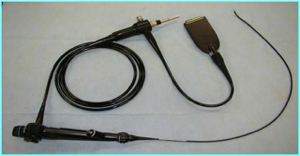
The “ureter” is the natural, narrow tube that drains each kidney. This is the tube where kidney stones may try to pass through and get stuck, causing pain. In the past 20 years, with the introduction of fiberoptic technology, we have developed thin cameras that are able to be placed in the ureter.
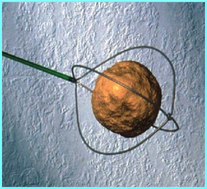
We are able to see inside the ureter and perform various procedures including the use of a small laser fiber to break stones into small fragments. We also have tiny wire baskets that we can use to grab small pieces of stones and remove them outside the body.
Almost all types of stones may be treated with ureteroscopy. Obviously, the larger the stone is, the longer it may take to break and remove each piece.
At the end of the procedure, most surgeons leave a stent (plastic tube) inside the ureter with a tip in the bladder. The stent is placed because the ureter gets swollen after instrumentation and the swelling may block the kidney after surgery (just like if you hit your finger, it gets swollen).
This surgery is normally done in the outpatient setting and thus patients are sent home same day unless there are other health problems that require an admission to the hospital.
What it involves?
After patients are asleep with general anesthesia, patients lay on their back in stirrups. The surgeon initially uses a small camera to look inside the bladder and place a small wire up to the kidney that has the kidney stone. Using the wire as our guide, surgeon places the thinner camera (ureteroscope) and may see the entire drainage tube (ureter) as well as the inside of the kidney.
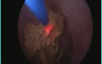
Once the stone is identified, a special tiny laser fiber is used to crack the stone into very fine fragments which then are left to pass naturally or if large enough are then removed with tiny baskets that are placed through the camera.
At the end of the procedure, most surgeons leave a stent to help the ureter recover from the surgery.
Length of Surgery
Ureteroscopy length of surgery primarily depends on the size of the stone. Most ureteroscopy procedures take about 1-2 hours to complete.
Anesthesia General anesthesia (heavy sedation with a ventilator) is recommended for best results. With general anesthesia, we are able to control patient’s breathing and thus allow for better targeting of the stone as we use the laser to break the stone.
Benefits
- Minimally-invasive approach
- Go home same day
- No skin incisions
- Works well with smaller stones
- Higher rate of complete stone clearance when compared to ESWL
- Lasers, unlike sound waves, are able to fragment almost every type of stone
Acute Risks
- Possible incomplete stone removal
- Urine infection requiring hospitalization
- Potential damage to the ureter or kidney
- Stent related pains, (bladder and/or kidney pains)
- Blood in the urine is very common and resolves within the first 24-48 hours, though as long as the stent is in place, some mild bleeding is very much expected
Chronic Risks
- Further stone production
- Potential for scar tissue development along the ureter that may produce obstruction of the kidney
