Broward Urology Center physicians see a lot of people in consultation regarding “kidney cysts.” We hope this page explains the basics about kidney cysts so that you may become more knowledgeable regarding this condition. Please feel free to ask us more details during your consultation visit.
What Are Kidney Cysts?
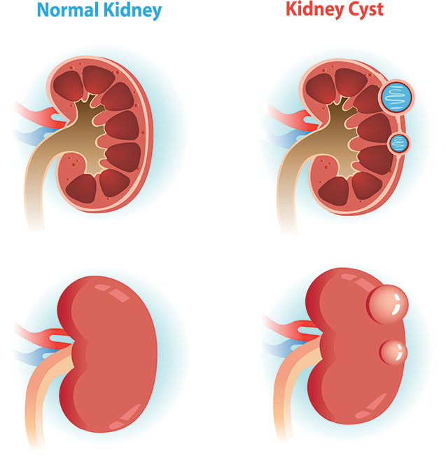
Cysts are fluid filled sacs that may occur in various parts of the body. They resemble a pimple or a boil. Certain organs tend to get cysts especially when those organs are made out of small tubes. For instance, the liver makes a fluid called bile to help you digest food and bile travels in many small tubes within the liver. The liver is very prone to cysts. Cysts are also commonly found in the testicles and organs that drain the testicles, the epididymis. Well, the kidney is made out of millions of tubes that filter blood and make urine. It is thought that some of these tubes may break and create these pockets of fluid that become a cyst.
Are Kidney Cysts Rare?
No, kidney cysts are very common and may be found in over 50% of people over 50 years of age.
What Causes Kidney Cysts?
For most part, kidney cysts are a random occurrence. However, there are certain genetic conditions and medical conditions that may cause kidney cysts. A rare hereditary condition called a “polycystic kidney disease” that runs in certain families causes hundreds of cysts to form in the kidneys. Patients on chronic dialysis are also more prone to kidney cysts.
There is nothing that a person does in their life that may cause kidney cysts. No lifestyle behavior, environmental exposure or diet has been associated with kidney cysts.
Are There Any Concerns with Kidney Cysts?
Most kidney cysts are not a concern as most cysts are what we call “simple cysts”. Simple cysts are cysts that only have clear fluid inside them and do not pose any risk. In rare cases, simple cyst may be problematic if they are large and are located in an area that may be obstructing the outflow of urine from the kidney. Sometimes, simple cysts may be large enough to cause some pain.
Rarely, however, there are cysts that are actually cancerous. The way we may distinguish the benign from cancerous (malignant) cysts is by way of imaging such as Kidney Ultrasound, CT scan or MRI.
Rarely, however, there are cysts that are actually cancerous. The way we may distinguish the benign from cancerous cysts is by way of imaging such as Kidney Ultrasound, CT scan or MRI.
How Can You Tell If a Kidney Cyst Is Cancerous?
On kidney ultrasound, if a cyst contains only some fluid inside of it and nothing else, it is typically a simple and benign cyst. If the cyst contains something inside besides fluid, we call these “complex cysts”.
The best way to judge whether a complex cyst is something to be concerned about is by way of CT or MRI pictures of the kidney. Once we look at those images with you, then we may be able to tell if a cyst is abnormal and judge whether the risk of a malignancy is high or low. You may hear us refer to a “Bosniak Classification” during your consultation. This is a way to categorize a cyst on a scale from 1 to 4 as to the risk of a malignancy for a particular cyst, based on how it appears on imaging.
How Do We Treat Kidney Cysts?
Treatment of kidney cyst depends on the risk of malignancy. We classify this on a Bosniak scale which is basically a way to group different cysts into different categories of risk.
Bosniak 1 Cyst
- this refers to a “simple cyst”
- the cyst is just filled with fluid
- risk of cancer is <1%
- what to do? Typically nothing. We don’t recommend follow up. However, if the cyst is large or in a location where it may block the urine flow if it becomes larger, it would be good to keep an eye on it with repeat imaging.
Bosniak 2 Cyst
- this type of cyst may have thin tissue inside of it dividing it (thin wall) OR it may have a small calcium deposit on the edge of the cyst OR the fluid inside the cyst appears thick
- risk of cancer <3%
- what to do? Typically nothing. Follow up is not recommended.
Bosniak 2F Cyst
- this cyst is similar to the Bosniak II cyst, however, the wall inside the cyst is thicker or the cyst is >3 cm in size
- risk of cancer 5-10%
- what to do? We recommend repeating imaging of the cyst over time to see if it changes. We use CT, MRI or Ultrasound pictures to follow cysts. Please click here to read more about the differences between the imaging tests.
Bosniak 3 Cyst
- these types of cysts are certainly a concern in our field. They are distinct from the above cysts in that the tissue inside the cyst actually appears to have what’s called “enhancement” which means that the tissue inside the cyst has blood flow. We can detect “enhancement” by comparing CT or MRI imaging before and after IV dye is given during the scan. If the area within the cyst “lights up” with the IV dye, that means the tissue inside is alive and thus the risk of cancer increases
- risk of cancer 40-60%
- what to do? Surgery to remove this cyst is the standard recommendation. In select cases, we keep a close eye on these cysts and repeat imaging tests to see if they change in size or character.
Bosniak 4 Cyst
- these types of cysts are considered to be tumors with some fluid inside of them. Imaging of these cysts typically shows thick, living tissue inside the cyst.
- risk of cancer is >80%
- we treat these types of cysts like any kidney cancer with surgical removal of either the cyst or the whole kidney depending on the size and location. Please see our Kidney Cancer page for more details.
